Shockwave Therapy for the Face and Neck: Skin Tightening, Tension Headaches, Neck Pain and Jaw Pain

Shockwave Therapy for the Face and Neck, Shockwave therapy — also known as acoustic wave therapy or extracorporeal shockwave therapy (ESWT) — is a non-invasive treatment that uses focused sound-pressure waves to switch on the body’s own healing.

In aesthetic medicine it is increasingly explored for facial skin tightening and rejuvenation. In sports and musculoskeletal medicine — the focus at Triaxis — the same technology offers a drug-free, needle-free way to ease the muscle-driven tension behind many tension headaches, neck pain and jaw pain.

nerve root irritation at the spine

What is shockwave therapy?

Shockwave therapy (ESWT) delivers high-energy acoustic (sound-pressure) waves to targeted tissue. These waves set off a cascade of biological responses — improved blood flow, the release of growth factors, and the calming of overactive pain signals — that together promote healing and relieve pain.

Originally developed to break down kidney stones, shockwave technology has become a cornerstone of modern musculoskeletal and sports medicine. It is entirely non-surgical, requires no anaesthetic and no downtime, and is delivered in short, comfortable sessions in the clinic.

Shockwave therapy in facial aesthetics

Acoustic wave therapy has become a genuine talking point in aesthetic medicine. Because the same sound-pressure waves stimulate fibroblasts — the cells that produce collagen and elastin — clinicians and researchers have explored whether the technology can firm and refresh facial skin without needles, surgery or heat.

The early findings are encouraging. In one study of 340 patients, focused and radial acoustic wave therapy produced measurable facial tightening confirmed on three-dimensional imaging, and the authors described it as a safe, non-invasive route to skin rejuvenation[6]. More recent work using ultra-pulse shockwave technology similarly reported improvements in skin texture and firmness[7]. Beyond firmness, it is reportedly used to soften fine lines and wrinkles and to help lift areas of lax skin[7].

This is why acoustic wave therapy for the face has drawn interest as a non-surgical facial rejuvenation option — a needle-free, no-downtime approach for people who would rather avoid injections or surgery. In aesthetic practice it is reportedly explored across a range of concerns:

Aesthetic focus What acoustic wave therapy is reportedly used for
Skin firmness & tightening Stimulating collagen and elastin to help firm lax or ageing skin
Fine lines & wrinkles Softening the appearance of expression lines and wrinkles
Facial contour & lifting Helping lift and redefine areas of sagging, such as along the jawline and cheeks
Skin texture & pores Refining overall skin texture and the look of enlarged pores
Non-surgical profile No needles, no incisions and no downtime; non-thermal, so suitable across skin types


Because the treatment is non-thermal, it does not rely on heat and is generally considered suitable for all skin types, including deeper skin tones. For anyone weighing up facial tightening or skin rejuvenation options, it offers a gentle, non-invasive route worth discussing with a doctor who can assess whether it fits your skin and goals.

From aesthetics to relief: why the same technology eases pain

Here is the link many people miss. The very tightness people notice in the face, jaw and neck is often muscular — and treating tight, painful muscle is exactly where shockwave therapy is at its strongest and best-evidenced.

It works through a process called mechanotransduction: cells sense the mechanical energy of the waves and convert it into biological signals that switch on repair[4]. In practical terms, three helpful things happen at once — blood flow rises and new microvessels form; the body releases growth factors while dialling down inflammatory chemicals; and overactive pain-signalling nerve endings are calmed[4]. This is why the same device explored for firming skin can also release the tight, tender knots in muscle known as myofascial trigger points.

Myofascial trigger points are hyper-irritable spots within a tight band of muscle. When pressed, they reproduce pain that spreads elsewhere — a trigger point in the neck can send pain into the head, and one in the jaw muscles can radiate across the face.

At a glance: how shockwave therapy helps facial and head pain

Condition What’s often behind it How shockwave therapy helps
Tension headaches Tight neck, shoulder and scalp muscles; trigger points Releases the trigger points that refer pain to the head[1]
Neck pain Upper trapezius and side-of-neck muscle tension Reduces pain and stiffness and improves function[2][3]
Jaw pain (TMD) Overworked masseter and temporalis chewing muscles Clinically meaningful pain relief and better mouth opening[4][5]


Shockwave therapy for tension headaches

Most tension-type headaches are not a problem inside the head at all — they are referred pain from tight muscles in the neck, shoulders and scalp. Because shockwave therapy targets exactly these overworked muscles and their trigger points, it offers a promising, drug-free route to relief.

In practice, settling the trigger points that feed a headache can ease symptoms that have resisted medication. A published case of chronic, treatment-resistant tension-type headache improved as part of a programme that included shockwave therapy, with a noticeable effect after treatment[1]. For anyone tired of reaching for painkillers, addressing the muscular source is a welcome alternative.

Shockwave therapy for neck pain

Neck pain and tension headaches often travel together, driven by the same tight muscles — particularly the upper trapezius and the muscles along the side of the neck. The evidence here is strong. In a randomised trial of patients whose headaches arose from neck trigger points, shockwave therapy reduced pain, improved function and reduced muscle stiffness — performing as well as hands-on manual therapy[2][3]. Relieving this muscular tension frequently eases both the neck pain and the headaches it refers.

Shockwave therapy for jaw pain (TMD)

Jaw pain, facial aching, difficulty opening the mouth and jaw clicking are hallmarks of temporomandibular disorders (TMD) — and much of this is muscular, centred on the powerful masseter and temporalis chewing muscles. This is one of the most exciting areas for shockwave therapy.

A 2026 meta-analysis pooling 18 randomised trials found moderate-to-high certainty evidence that shockwave therapy produces clinically meaningful reductions in jaw pain and improvements in mouth opening[4]. A separate randomised trial applying shockwave to the masseter reported effective pain relief with no major side effects[5]. For the many people whose jaw tension also drives facial pain and headaches, that is genuinely good news.

What a session involves

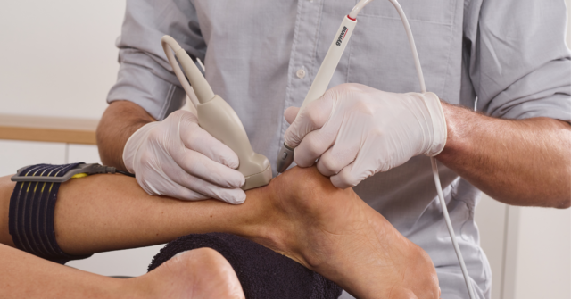
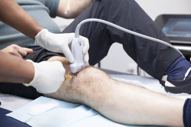
A shockwave session is quick and straightforward. After a clinical assessment — supported by ultrasound examination where helpful — the treatment head is applied to the target muscle with a conducting gel. Most people feel a firm tapping sensation, and the intensity is tailored to your comfort. There are no needles, no anaesthetic and no downtime, so you can return to your day straight away.

What to expect Shockwave therapy at Triaxis
Anaesthetic needed No
Needles No
Downtime None — resume normal activity immediately
Typical session length Around 10–20 minutes
Typical course Several sessions, usually about a week apart
Delivered by Doctor-led, with ultrasound guidance where useful


Is shockwave therapy safe? Who is it for?

Shockwave therapy has a strong safety record. Across the studies above, side effects were minor and short-lived — usually mild, temporary tenderness or redness at the treated area — with no major complications reported[4][5]. Because it is non-thermal, it is also suitable across all skin types.

As with any treatment, it is not for everyone. It is generally avoided over areas of active infection, over tumours, in people with certain bleeding disorders or taking blood-thinning medication, and during pregnancy. A proper medical assessment confirms it is the right choice for you — one of the real advantages of being treated in a doctor-led clinic.

Frequently asked questions on Shockwave Therapy for the Face and Neck

Can shockwave therapy be used as part of a facial aesthetics or rejuvenation programme?

Yes — acoustic wave therapy is increasingly used in aesthetic medicine for non-surgical facial tightening and skin rejuvenation, and it can sit alongside other treatments. A consultation confirms what suits your skin and goals.

Is facial shockwave therapy surgical, and is there downtime?

No. It is non-surgical and non-invasive, with no needles and no downtime, so you can return to your normal day straight away.

Does shockwave therapy hurt?

Most people describe a firm tapping sensation rather than pain, and the intensity is adjusted to your comfort throughout.

How many sessions will I need?

It varies with your condition, but a typical course runs over several weekly sessions. Your doctor will advise after assessing you.

Is there any downtime?

No. You can return to your normal activities immediately after a session.

Can it replace my headache medication?

It targets the muscular source of many tension headaches and can reduce reliance on painkillers, but any change to medication should be discussed with your doctor.

Why choose Triaxis

At Triaxis Sports & Joint Clinic, shockwave therapy is delivered by Dr Dinesh Sirisena, a Consultant in Sports, Exercise and Musculoskeletal Medicine with experience at the highest levels of elite sport. Every treatment begins with a careful diagnosis — often supported by ultrasound imaging — so the therapy is aimed precisely where it will help most. Whether your concern is a nagging tension headache, a stiff and painful neck, or an aching jaw, you are in expert, doctor-led hands.

Ready to find lasting relief? Book a consultation at Triaxis Sports & Joint Clinic, Farrer Park Hospital.
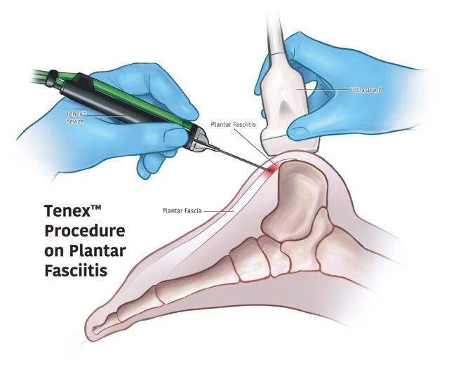
Tenex Therapy: Uses, Evidence, Risks and What to Expect

Written and medically reviewed by
Dr Dinesh Sirisena
Consultant in Sports, Exercise & Musculoskeletal Medicine
Triaxis Sports & Joint Clinic

Tenex therapy is a minimally invasive procedure that uses ultrasound guidance and ultrasonic energy to remove selected abnormal soft tissue through a small skin opening. It is mainly used for persistent tendon and fascia conditions that have not improved with appropriate conservative care. Studies report improvements in pain and function in selected patients, but much of the available evidence is based on observational studies rather than large comparative trials.

Tenex Therapy

What is Tenex therapy?

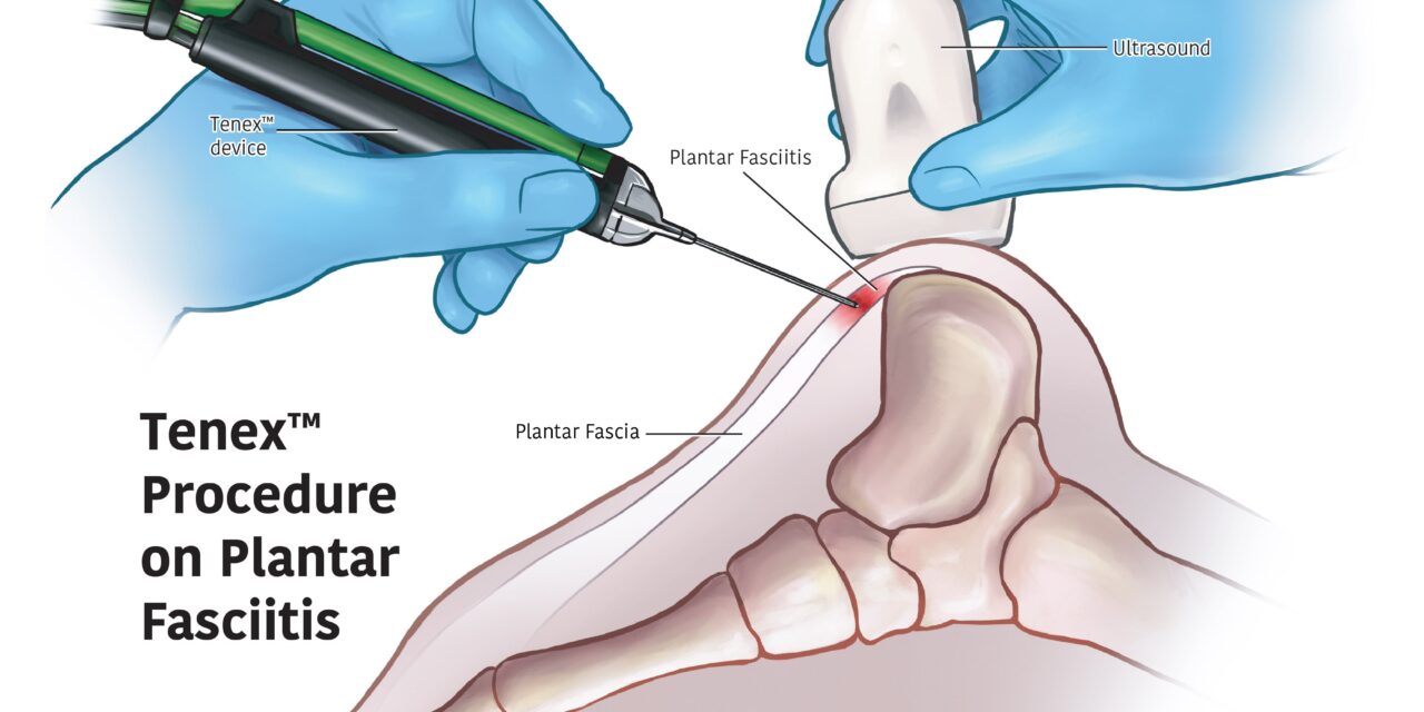
Tenex therapy is a minimally invasive procedure used to remove selected abnormal tendon or fascia tissue through a small skin opening. The Tenex Health TX system uses ultrasonic energy together with irrigation and aspiration, while imaging helps the clinician identify and treat the intended area.

The formal medical terms are usually:

  • Percutaneous ultrasonic tenotomy for tendon procedures
  • Percutaneous fasciotomy for fascia procedures

The Tenex Health TX system is classified by the U.S. FDA as an ultrasonic surgical instrument. FDA records describe it as an ultrasonic surgical aspirator that fragments, emulsifies and removes soft tissue.

How does Tenex therapy work?

Tenex uses a small ultrasonic instrument placed into the treatment area under image guidance. The device delivers ultrasonic energy to fragment selected tissue while fluid irrigates the area and aspiration removes the fragmented material. The aim is to remove targeted abnormal tissue rather than simply inject medication into it.

The basic process involves three functions:

Ultrasonic energy
The device tip vibrates at ultrasonic frequency and interacts with the selected tissue.

Irrigation
Fluid is delivered to the treatment area.

Aspiration
Fragmented tissue and fluid are removed through the device.

This makes Tenex different from steroid injections, prolotherapy or PRP.

Tenex works by removing selected tissue that is not healing. Recovery is then supported by the body’s natural healing, rehabilitation and a gradual return of tendon capacity.

What conditions has Tenex therapy been studied for?

Tenex has primarily been studied for chronic tendinopathy and selected fascia conditions that remain symptomatic despite conservative treatment. Published studies include several tendon regions, although the amount and quality of evidence vary significantly between anatomical sites.

Research has included:

  • Tennis elbow or lateral elbow tendinopathy
  • Golfer’s elbow or medial elbow tendinopathy
  • Achilles tendinopathy
  • Patellar tendinopathy
  • Gluteal tendinopathy
  • Rotator cuff-related tendon disease
  • Plantar fasciopathy
  • Other selected chronic tendon disorders

A 2024 systematic review identified studies across 11 tendon sites and found that published reports generally showed improvements in pain and function after percutaneous needle tenotomy or Tenex.

This is why results from one tendon should not automatically be applied to another.

What is tendinopathy and why can it become persistent?

Tendinopathy is a disorder of a tendon that can cause pain, reduced function and difficulty tolerating physical load. It commonly develops after repeated loading, sudden changes in activity or a combination of mechanical and individual factors. Persistent tendinopathy is more complex than simple inflammation.

Tendons connect muscles to bones and transfer force during movement.

For example:

  • The Achilles tendon transfers calf muscle force during running and jumping.
  • The patellar tendon helps transmit force through the knee.
  • The common extensor tendon at the elbow is heavily loaded during gripping.
  • The gluteal tendons help stabilise the hip during walking and running.

Persistent tendon symptoms may be associated with changes in tendon structure and load tolerance.

Therefore, treatment usually needs to address more than pain alone.

Who may be considered for Tenex therapy?

Tenex may be considered in selected patients with persistent tendon or fascia symptoms when the diagnosis is reasonably clear and appropriate conservative management has not provided sufficient improvement. It is not normally a first-line procedure for every episode of tendon pain.

Clinical assessment may consider:

  • Duration of symptoms
  • Exact anatomical diagnosis
  • Severity of pain and functional limitation
  • Previous rehabilitation
  • Exercise and tendon-loading history
  • Occupational demands
  • Sporting requirements
  • Relevant imaging
  • Other medical conditions
  • Alternative treatment options

A person with six weeks of tendon pain may therefore be managed very differently from someone with longstanding symptoms despite structured rehabilitation.

Does Tenex therapy actually work?

Studies of Tenex and similar percutaneous ultrasonic tenotomy procedures report improvements in pain and function for many patients with persistent tendinopathy. As a newer procedure, it continues to be studied, with larger comparative trials ongoing.

A 2021 systematic review found promising results for percutaneous ultrasonic tenotomy across a range of tendon conditions.

A newer 2024 systematic review included 10 studies covering 11 tendon sites. It reported improvements in:

  • Pain
  • Function
  • Quality of life

with relatively few reported adverse effects.

For most patients, Tenex is best considered as one option within a broader plan for persistent tendon pain.

Is Tenex the same as tendon surgery?

Tenex is a percutaneous surgical procedure, but it differs from conventional open tendon surgery. It is performed through a small skin opening and uses ultrasonic instrumentation rather than a larger surgical exposure. The exact procedure, anaesthesia and recovery plan depend on the tendon and amount of tissue involved.

It is sometimes described as “non-surgical,” and Tenex themselves describe their underlying procedures as percutaneous tenotomy or fasciotomy.

Therefore, a more accurate patient description is:

“A minimally invasive percutaneous procedure” — i.e. treating a tendon through a small puncture in the skin rather than open surgery.

How is Tenex different from steroid injections, PRP and prolotherapy?

Tenex physically removes selected tissue using ultrasonic instrumentation, whereas steroid injections, PRP and prolotherapy involve introducing substances into or around musculoskeletal structures. Their mechanisms, evidence and clinical uses are different, and none should be assumed to be universally superior.

Steroid injection
Uses corticosteroid medication to reduce inflammation in selected conditions.

PRP
Uses a platelet-containing preparation derived from the patient’s blood.

Prolotherapy
Commonly uses a dextrose-containing solution.

Tenex
Uses ultrasonic instrumentation to fragment and remove selected tissue.

The choice, if any, should depend on the diagnosis and evidence for the specific tendon problem rather than simply choosing the newest procedure.

Why is musculoskeletal ultrasound used with Tenex?

Musculoskeletal ultrasound can help identify the affected tendon or fascia, assess surrounding structures and guide the Tenex instrument toward the intended area during the procedure. Ultrasound provides real-time imaging, but it does not by itself determine whether Tenex is necessary or guarantee a successful outcome.

Ultrasound may help assess:

  • Tendon thickness
  • Tendon structure
  • Partial tears
  • Calcification
  • Surrounding bursae
  • Nearby nerves and blood vessels
  • Plantar fascia

During the procedure, imaging helps the clinician monitor the instrument relative to the target anatomy.

However, an abnormal ultrasound image does not automatically mean tissue should be removed.

Clinical symptoms and examination findings remain important.

Do I need an MRI before Tenex therapy?

Not everyone being assessed for Tenex needs an MRI. The appropriate imaging depends on the tendon, symptoms and clinical question. Musculoskeletal ultrasound is commonly useful because it can assess many superficial tendons dynamically, while MRI may be considered when deeper anatomy or another diagnosis needs clarification.

Possible imaging includes:

X-ray
May help identify calcification, bone spurs or other bone-related findings.

Musculoskeletal ultrasound
Can assess many tendons and guide the procedure in real time.

MRI
May be useful for deeper structures, more complex injuries or when another diagnosis is suspected.

Imaging should be used when it helps answer a clinical question, rather than as an automatic prerequisite.

What happens during a Tenex procedure?

During a Tenex procedure, the clinician identifies the treatment area using imaging, prepares the skin and administers anaesthesia as appropriate. A small opening is made, and the ultrasonic instrument is introduced toward the selected tissue under image guidance. The device then fragments and removes targeted tissue using ultrasonic energy, irrigation and aspiration.

A typical process may include:

  1. Review of the diagnosis and treatment indication.
  2. Ultrasound assessment of the tendon or fascia.
  3. Discussion of benefits, limitations and risks.
  4. Skin preparation and local anaesthesia.
  5. Small skin opening.
  6. Image-guided placement of the instrument.
  7. Ultrasonic treatment and removal of selected tissue.
  8. Dressing of the treatment site.
  9. Post-procedure instructions.
  10. Rehabilitation planning.

The exact steps vary according to the anatomical area.

Is Tenex therapy painful?

Tenex is commonly performed with local anaesthesia, so discomfort may be reduced during the procedure. However, patients may still experience pressure, soreness or discomfort during recovery. The amount varies with the treatment site, amount of tissue treated and individual response.

The manufacturer describes local anaesthesia as commonly used during the procedure.

After treatment, temporary symptoms may include:

  • Local soreness
  • Tenderness
  • Mild swelling
  • Bruising
  • Discomfort with movement

Patients should receive clear advice about what to expect and when symptoms warrant further medical assessment.

The procedure should not be advertised as “pain-free”.

What are the possible risks of Tenex therapy?

As a percutaneous procedure, Tenex carries potential risks such as temporary pain, bleeding, bruising, infection and injury to surrounding structures. The exact risks depend on the anatomical area and individual medical circumstances. Published studies generally report few complications, but the available evidence is still limited.

Potential considerations include:

  • Bleeding
  • Bruising
  • Infection
  • Temporary pain flare
  • Irritation of surrounding tissues
  • Nerve or blood vessel injury
  • Incomplete improvement
  • Persistent or recurrent symptoms

Patients should also tell the clinician about:

  • Blood-thinning medication
  • Diabetes
  • Allergies
  • Active infection
  • Previous procedures
  • Relevant medical conditions

No procedure should be described as risk-free.

How long does recovery take after Tenex?

Recovery varies according to the tendon or fascia treated, severity of the original condition, physical demands and rehabilitation plan. Some activities may be restricted initially, followed by progressive movement and strengthening. A fixed return-to-sport timeline should not be promised because tendon recovery varies between individuals.

The manufacturer describes recovery instructions as varying by condition and notes that range-of-motion and strengthening exercises may be introduced after an initial period of rest.

A sensible rehabilitation plan may assess:

  • Pain during daily activities
  • Range of motion
  • Strength
  • Tendon load tolerance
  • Work requirements
  • Running or jumping ability
  • Sport-specific function

Return to activity should therefore be based on progress, not simply the number of weeks since the procedure.

Do I still need rehabilitation after Tenex?

Yes, rehabilitation may remain important because removing selected tendon tissue does not automatically restore strength, movement or load tolerance. Progressive exercise can help the affected tendon and surrounding muscles adapt to everyday, occupational and sporting demands during recovery.

Rehabilitation may include:

  • Education
  • Temporary activity modification
  • Range-of-motion exercises
  • Progressive strengthening
  • Tendon loading
  • Movement retraining
  • Return-to-running progression
  • Sport-specific exercises

The exact programme depends on the anatomical site.

For example, Achilles rehabilitation differs substantially from elbow or gluteal tendon rehabilitation.

Tenex should therefore be viewed as one part of a broader management pathway where appropriate.

Can athletes have Tenex therapy?

Tenex may be considered for selected athletes with persistent tendinopathy, but sporting participation alone is not a reason to undergo the procedure. Diagnosis, previous rehabilitation, tendon function, competition demands and the available evidence should guide whether an intervention is appropriate.

Relevant sports may include:

  • Running
  • Tennis
  • Golf
  • Badminton
  • Football
  • Rugby
  • Swimming
  • Cricket
  • Weightlifting

Different sports also load different tendons.

A runner with Achilles tendinopathy, for example, requires different assessment and rehabilitation from a tennis player with lateral elbow tendinopathy.

The objective should be restoring appropriate function rather than simply reducing pain quickly.

When should I seek assessment for persistent tendon pain?

Consider medical assessment when tendon pain persists, repeatedly returns or limits work, exercise or sport despite appropriate load modification and rehabilitation. Prompt assessment is particularly important after sudden injury, significant weakness, inability to bear weight or concern about a tendon rupture.

Assessment may be helpful when there is:

  • Persistent pain over several weeks or months
  • Reduced strength
  • Repeated flare-ups
  • Difficulty returning to sport
  • Failure to progress with rehabilitation
  • Sudden loss of function
  • Significant swelling or bruising
  • Concern about a partial or complete tendon tear

The first priority is establishing the diagnosis rather than deciding on a procedure.

How is Tenex therapy considered at Triaxis Sports & Joint Clinic?

At Triaxis Sports & Joint Clinic, Tenex may be considered for selected persistent tendon or fascia conditions following Sports Medicine and musculoskeletal assessment. The decision depends on the diagnosis, previous rehabilitation, functional limitations, imaging where appropriate, available evidence and alternative management options.

Assessment may include:

  • Sports Medicine evaluation
  • Musculoskeletal examination
  • Sports ultrasound where appropriate
  • Review of previous imaging
  • Assessment of strength and tendon loading
  • Review of rehabilitation already attempted
  • Patient education
  • Rehabilitation planning
  • Shared decision-making

Where Tenex is considered, patients can discuss:

  • Why the procedure is being considered
  • Which structure is being treated
  • Current evidence
  • Potential risks
  • Recovery expectations
  • Rehabilitation afterwards
  • Other treatment options

Depending on the individual’s condition and clinical assessment, certain procedures may be considered as part of a broader management plan.

Singapore MOH requires healthcare advertising content to remain accurate and truthful and not contain exaggerated claims that could encourage unwarranted healthcare use.

Frequently Asked Questions About Tenex Therapy

Does Tenex therapy cure tendinopathy?

No. Tenex should not be described as curing tendinopathy. It uses ultrasonic instrumentation to remove selected tissue, but pain and function depend on multiple factors including tendon load, strength, rehabilitation and the underlying condition. Current studies report improvements in selected patients, but the evidence base remains largely observational and does not establish guaranteed or permanent results.

Is Tenex therapy FDA approved?

A more accurate term is FDA cleared, rather than “FDA approved.” The Tenex Health TX System has received U.S. FDA 510(k) clearance as an ultrasonic surgical instrument. FDA records describe the system as an ultrasonic surgical aspirator. Clearance of the device does not mean Tenex has been proven superior to other treatment options for every tendon disorder.

Is Tenex better than open surgery?

Tenex is less invasive because it uses a small percutaneous approach, but surgical decisions depend on the type and severity of tendon pathology, previous treatment and individual needs. Some conditions may not require surgery at all, while others may require conventional surgical assessment.

Can Tenex treat tennis elbow?

Tenex has been studied for chronic lateral elbow tendinopathy, commonly called tennis elbow, and published observational studies have reported improvements in selected patients. However, exercise-based rehabilitation remains an important management option, and Tenex should be undertaken as part of a combined treatment plan.

Can Tenex be used for Achilles tendinopathy?

Percutaneous ultrasonic tenotomy has been studied for Achilles tendinopathy. Not all Achilles problems are suitable for the procedure. Insertional tendinopathy, mid-portion tendinopathy, partial tearing and other causes of posterior ankle pain may require different management.

Does Tenex remove healthy tendon?

The intention is to identify and remove selected abnormal tissue while preserving surrounding structures, using imaging to guide the procedure. The procedure still depends on appropriate diagnosis, imaging interpretation and clinical technique.

How soon can I return to sport after Tenex?

There is no universal return-to-sport timeline. Progress depends on the tendon treated, symptoms, strength, sporting demands and rehabilitation. Patients may need a period of activity modification followed by progressive strengthening and gradual reintroduction of sport-specific loads. A fixed promise such as returning to sport within a particular number of weeks would not be appropriate.

Can Tenex replace physiotherapy or rehabilitation?

Not necessarily. Tenex addresses selected tissue but does not automatically restore strength, movement or tendon capacity. Rehabilitation may remain important before and after the procedure. Depending on the tendon involved, this may include progressive strengthening, load management, mobility work and sport-specific rehabilitation.

About the Author

Dr Dinesh Sirisena is a Consultant in Sports, Exercise & Musculoskeletal Medicine. His clinical work includes assessment and management of sports injuries and musculoskeletal conditions affecting tendons, muscles, joints, ligaments and related structures.

Medical Disclaimer

This page provides general educational information and does not replace individual medical advice. Tenex therapy is not necessary or appropriate for every tendon or fascia condition. Decisions about imaging, rehabilitation or percutaneous procedures should be based on individual symptoms, diagnosis, medical history, clinical findings and treatment goals.

Ultrasound-Guided Hydrodissection: Uses, Evidence and What to Expect

Written and medically reviewed by
Dr Dinesh Sirisena
Consultant in Sports, Exercise & Musculoskeletal Medicine
Triaxis Sports & Joint Clinic

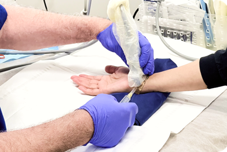
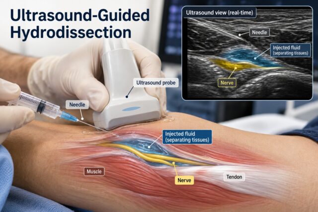
Hydrodissection is a minimally invasive, ultrasound-guided procedure that uses fluid to gently separate tissues and free nerves, fascia and tendons from the structures pressing on or sticking to them.

It is used across a broad range of musculoskeletal problems — including nerve entrapments such as carpal tunnel syndrome, fascial and myofascial pain, and persistent tendon conditions such as Achilles and patellar tendinopathy. The technique is performed under real-time ultrasound so the clinician can see the needle and the injected fluid throughout, and it works well alongside rehabilitation and progressive loading.

Ultrasound-Guided Hydrodissection

What is hydrodissection?

Hydrodissection is an ultrasound-guided injection technique in which fluid is used to create space around a nerve, fascial layer or tendon, gently separating it from the surrounding tissue. This can relieve compression, reduce adhesions and improve the way the structure glides and moves.

Because it is performed under high-resolution ultrasound, the clinician can identify the exact tissue plane and watch the fluid spread in real time. The technique is also described in the literature as perineural injection therapy when applied to nerves, and as high-volume injection or high-volume image-guided injection (HVIGI) when a larger fluid volume is used around a tendon.

Hydrodissection describes the technique, not a single medicine. Depending on the problem, the injected solution may include:

  • Normal saline
  • Local anaesthetic
  • 5% dextrose (D5W), often used for nerve-related pain
  • Platelet-rich plasma (PRP) in selected cases
  • Corticosteroid, where clinically appropriate

The choice of solution is tailored to the diagnosis, the structure being treated and the individual patient.

What conditions can hydrodissection treat?

Hydrodissection is used across three broad areas: freeing entrapped nerves, releasing adhered fascial and myofascial planes, and addressing the tissue changes around persistent tendon problems. The strongest evidence is for nerve entrapment, while its use around fascia and tendons continues to grow.

Nerve entrapment (nerve hydrodissection)

This is the best-established application. Fluid is used to release a compressed nerve from the tissue trapping it, restoring space and improving nerve gliding. Commonly treated entrapments include:

  • Carpal tunnel syndrome (median nerve at the wrist)
  • Cubital tunnel syndrome (ulnar nerve at the elbow)
  • Deep gluteal / sciatic nerve entrapment
  • Meralgia paraesthetica (lateral femoral cutaneous nerve)
  • Baxter’s nerve and tarsal tunnel entrapment in the heel and foot
Fascial and myofascial release

Fluid can be used to separate adhered fascial layers and interfascial planes that contribute to pain and restricted movement, for example around the plantar fascia or the gluteal and piriformis region. This is an active and growing area of practice.

Tendon and paratendon problems (high-volume injection)

Around persistent tendon conditions such as mid-portion Achilles and patellar tendinopathy, a higher volume of fluid is placed between the tendon and the tissue in front of it — sometimes called high-volume “stripping.” The aim is to influence the small blood vessels and accompanying nerve fibres that are associated with tendon pain, as part of a wider loading and rehabilitation plan.

How does hydrodissection work?

Hydrodissection works mainly by mechanically separating structures that have become compressed or stuck together. Freeing a nerve from surrounding tissue can relieve pressure, improve its blood supply and restore normal gliding, while separating tissue planes around a tendon can influence the vessels and nerve fibres linked to pain.

For nerves, the goal is to release the nerve from compressive or scarred tissue so that its own small blood vessels and nerve fibres (the vasa nervorum and nervi nervorum) can function normally again. Some solutions, such as 5% dextrose, may also have a settling effect on irritated nerves beyond the mechanical release itself.

For tendons, chronic tendon pain is often accompanied by increased small blood vessels and nerve fibres around the tendon, which can be seen on Doppler ultrasound. Placing fluid between the tendon and the tissue in front of it is intended to separate these planes and influence those pain-associated structures. This is why the technique is combined with progressive tendon loading, which builds the tendon’s capacity over time.

What does the evidence show?

The evidence is strongest for nerve hydrodissection, where randomised trials and systematic reviews support its use in conditions such as carpal tunnel syndrome. Evidence for fascial applications is emerging, while for tendon conditions hydrodissection is best considered as one component of a rehabilitation-based programme.

For nerve entrapment, randomised controlled trials have shown that ultrasound-guided hydrodissection can improve symptoms and function in carpal tunnel syndrome, with benefits maintained over months. Systematic reviews and a network meta-analysis have reported that 5% dextrose is among the most effective injectates, with results comparable to corticosteroid at longer follow-up and an excellent safety profile. Contemporary reviews now position hydrodissection as a valuable step for patients who have not settled with splinting or therapy but who wish to avoid, or are not yet ready for, surgery.

For fascial and myofascial pain, published case series and ongoing trials describe encouraging results, and interest in interfascial hydrodissection is growing as ultrasound techniques improve.

For tendon conditions, results are more variable. A 2020 randomised controlled trial found that high-volume injection without corticosteroid did not add benefit over a small placebo injection when both groups followed a structured exercise programme, while a 2024 systematic review reported possible short-term improvement, particularly in protocols that included corticosteroid. Taken together, this supports using high-volume injection selectively and always alongside a progressive loading programme, rather than as a stand-alone treatment.

Is hydrodissection the same as prolotherapy, PRP or a steroid injection?

No. Hydrodissection describes an injection technique — using fluid under ultrasound to separate tissues — while prolotherapy, PRP and corticosteroid refer to the substances injected. Some hydrodissection procedures use one of these solutions, but the terms are not interchangeable.

In practice, the diagnosis and the structure being treated guide both the technique and the choice of solution. For example, a nerve entrapment and a chronic tendon problem may both be treated with hydrodissection, but with different injectates, volumes and target planes.

Why is ultrasound guidance used?

Ultrasound guidance lets the clinician see the nerve, fascia or tendon and the surrounding anatomy in real time, guide the needle precisely into the intended tissue plane, and watch the fluid spread exactly where it is needed. This precision is central to how hydrodissection works.

Sports and musculoskeletal ultrasound can also assess the target structure before treatment — for example the size and shape of a nerve, the thickness and structure of a tendon, or blood flow using Doppler imaging — and can be used dynamically while the joint or limb moves.

Who may be considered for hydrodissection?

Hydrodissection may be considered for people with a confirmed nerve entrapment, fascial pain or persistent tendon problem who are looking for a minimally invasive option, often after an appropriate period of conservative care. A clinical assessment, and usually ultrasound, help confirm the diagnosis and identify the right target.

Assessment typically considers:

  • The specific diagnosis and where the problem is arising
  • Duration and pattern of symptoms
  • Response to previous treatment and rehabilitation
  • Functional and sporting demands
  • Relevant medical conditions and current medications
  • Ultrasound findings

What happens during a hydrodissection procedure?

During hydrodissection, the area is examined with ultrasound, the skin is cleaned, and a needle is guided precisely toward the target nerve, fascia or tendon. Fluid is then introduced to separate the tissue planes while the clinician watches in real time. It is usually a day procedure.

A typical procedure involves:

  1. Clinical review and confirmation of the diagnosis
  2. Ultrasound examination of the target structure
  3. Discussion of the expected benefits, the solution used and any risks
  4. Skin preparation
  5. Ultrasound-guided needle placement
  6. Introduction of the tailored solution to separate the tissue planes
  7. Post-procedure advice and rehabilitation planning

Is hydrodissection painful?

Most people tolerate hydrodissection well. It involves a needle and the introduction of fluid, so some pressure or temporary soreness is normal, and local anaesthetic is often included to keep the procedure comfortable. The amount of discomfort varies from person to person.

Any discomfort is usually related to the needle, the pressure of the fluid or an already sensitive area, and tends to settle over the following days. You will be given individual advice on what to expect and how to look after the area afterwards.

What are the possible risks?

Hydrodissection has a strong safety profile, and studies of nerve hydrodissection have reported very few complications. As with any needle-based procedure, considerations can include temporary soreness, bruising, a small risk of bleeding or infection, and irritation of nearby structures. Additional considerations apply when medicines such as corticosteroid are included.

To keep the procedure as safe as possible, it is helpful to discuss beforehand:

  • Current and blood-thinning medication
  • Allergies
  • Diabetes
  • Previous injections or procedures in the area
  • Other relevant medical conditions

How long does recovery take, and will I still need rehabilitation?

Recovery varies with the condition treated and your individual response, and many people return to everyday activities quickly. Rehabilitation remains an important partner to hydrodissection — especially for tendon problems — because progressive loading builds lasting strength and capacity that an injection alone does not provide.

For nerve entrapments, symptom relief may develop over the weeks following the procedure. For tendon conditions, hydrodissection is most effective when combined with a graded loading programme, and return to running or sport is guided by function and comfort rather than a fixed timetable. Your clinician and rehabilitation team will tailor the plan to you.

How is hydrodissection considered at Triaxis Sports & Joint Clinic?

At Triaxis Sports & Joint Clinic, hydrodissection is considered following a Sports Medicine and musculoskeletal assessment. The clinic reviews the diagnosis, previous treatment, functional needs, ultrasound findings and the current evidence before discussing whether an ultrasound-guided procedure is a suitable option.

Where a procedure is appropriate, patients can discuss the solution being used, the expected benefits, any limitations and risks, and the alternatives, so that the decision is a shared one. Depending on the individual’s condition and clinical assessment, hydrodissection may be considered as part of a broader management plan that includes rehabilitation.

Frequently Asked Questions About Hydrodissection

Does hydrodissection help carpal tunnel syndrome?

Yes — this is one of its best-supported uses. Randomised trials and systematic reviews have shown that ultrasound-guided hydrodissection can improve symptoms and function in carpal tunnel syndrome, and it is increasingly offered to people who have not settled with splinting or therapy but wish to avoid or delay surgery. Your suitability is confirmed with a clinical assessment and ultrasound.

What is high-volume injection for tendons?

High-volume injection is the tendon-focused form of hydrodissection. A larger volume of fluid is placed between a painful tendon, such as the Achilles, and the tissue in front of it, to separate the planes and influence the small vessels and nerve fibres associated with tendon pain. It is used selectively and works best alongside a progressive loading programme.

Is hydrodissection a type of surgery?

No. Hydrodissection is a minimally invasive, ultrasound-guided injection performed as a day procedure. For conditions such as nerve entrapment, it is often considered as a step before surgery is needed.

What fluid is injected during hydrodissection?

It depends on the problem. Solutions may include saline, local anaesthetic, 5% dextrose, PRP or, where appropriate, corticosteroid. The choice is tailored to the diagnosis and the structure being treated, and you will be told what is being used.

Do I still need rehabilitation after hydrodissection?

For tendon conditions, yes — progressive loading builds lasting strength and capacity that an injection alone does not provide, so the two work best together. For nerve entrapments, activity and nerve-gliding advice often support recovery. Your plan is tailored to your condition.

How soon will I notice a difference?

This varies with the condition and the individual. Some people notice change within days, while for others relief develops over the following weeks. Return to sport is guided by function and comfort rather than a set number of days.

About the Author

Dr Dinesh Sirisena is a Consultant in Sports, Exercise & Musculoskeletal Medicine. His clinical work includes the assessment and management of sports injuries and musculoskeletal conditions involving muscles, tendons, ligaments, nerves, joints and related structures, including ultrasound-guided procedures.

Medical Disclaimer

This page provides general educational information and does not replace individual medical advice. Hydrodissection is not required or suitable for every person, and the right option depends on the diagnosis and clinical assessment. Decisions about imaging, rehabilitation or image-guided procedures should follow an individual consultation.

Hydrodilatation: Uses, Evidence, Risks and What to Expect

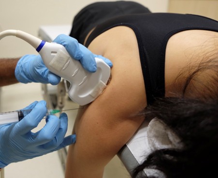
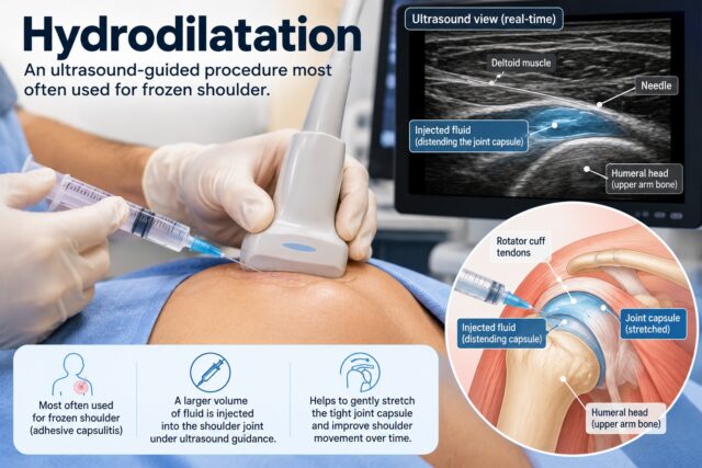
Hydrodilatation is an ultrasound-guided procedure most often used for frozen shoulder. A controlled volume of fluid introduced into the shoulder joint to gently expand the tightened joint capsule, usually together with local anaesthetic and, where appropriate, corticosteroid. It’s a well-tolerated day procedure with a strong safety record. It can help ease pain and improve movement — particularly external rotation, one of the movements most affected in frozen shoulder — and works best as part of a structured plan that includes rehabilitation.

Hydrodilatation is an ultrasound-guided shoulder procedure

What is hydrodilatation?

Hydrodilatation an ultrasound-guided shoulder procedure used most often for adhesive capsulitis, commonly called frozen shoulder. A controlled volume of fluid introduced into the glenohumeral (shoulder) joint to gently expand the tightened joint capsule. The fluid usually includes saline and local anaesthetic, with corticosteroid added where appropriate.

In frozen shoulder, the capsule surrounding the joint becomes tight, which brings on pain and progressively restricts movement. Hydrodilatation works with this problem directly: by introducing fluid under gentle pressure, it distends the capsule, creates more space within the joint, and helps restore movement.

Hydrodilatation also known as:

  • Hydrodistension
  • Joint distension
  • Capsular distension
  • Shoulder hydrodilatation

Although frozen shoulder the most common reason for the procedure, the same principle — using fluid to distend a tightened joint capsule under ultrasound guidance — can also be applied to help stiffness in other joints where the capsule has become restricted.

How does hydrodilatation work?

Hydrodilatation gently stretches and expands the tightened shoulder capsule by introducing fluid into the joint. This helps to create more room within the joint, ease movement and reduce pain. It’s a controlled, gradual process rather than a forceful one.

The shoulder joint is surrounded by a flexible sleeve of connective tissue called the joint capsule. In frozen shoulder, this capsule becomes tight and inflamed, and movement becomes restricted and uncomfortable.

During hydrodilatation, fluid is introduced into the glenohumeral joint under ultrasound guidance. The volume and gentle pressure distend the capsule, increase the available space within the joint, and help the shoulder move more freely. Where corticosteroid included, it can also help settle the inflammation that contributes to pain and stiffness.

This is best understood as gently distending and easing the capsule — creating room and improving mobility — rather than “breaking” or “tearing” anything. That description is both more accurate and a better reflection of what patients actually experience.

What’s frozen shoulder and why does the shoulder become stiff?

Frozen shoulder, or adhesive capsulitis, a condition in which shoulder pain and stiffness develop gradually because the capsule surrounding the joint becomes tight. Movement becomes restricted both when the patient moves the arm themselves and when a clinician moves the shoulder during examination.

The shoulder a ball-and-socket joint. The structures involved include:

Humeral head — the ball at the top of the upper arm bone.

Glenoid — the shallow socket on the shoulder blade.

Joint capsule — a flexible layer surrounding the joint.

Rotator cuff tendons — tendons that help control and stabilise shoulder movement.

Ligaments — connective tissues that help support the joint.

Muscles — including the rotator cuff, deltoid and shoulder blade muscles.

Nerves — supplying sensation and muscle control around the shoulder and arm.

In frozen shoulder, the main issue tightening of the joint capsule rather than simple muscle weakness — which exactly why a treatment aimed at the capsule itself, such as hydrodilatation, can be helpful.

Who may develop frozen shoulder?

Frozen shoulder commonly affects adults in middle age and can occur without any obvious injury. Certain medical conditions, particularly diabetes, are associated with a higher likelihood of developing it. A period of shoulder immobilisation after injury or surgery can also contribute.

Factors associated with frozen shoulder include:

  • Age, particularly middle age
  • Diabetes
  • Thyroid disorders
  • Previous shoulder injury
  • Shoulder surgery
  • Prolonged restriction of shoulder movement

Many people develop frozen shoulder without any clear trigger. Sports such as swimming, tennis, badminton, golf and weightlifting can make shoulder stiffness more noticeable, although the sport itself not usually the cause.

When may hydrodilatation be considered?

Hydrodilatation may be considered for patients with frozen shoulder when pain and stiffness continue to affect daily activity. It is one of several effective options and can be a helpful step for patients who want to ease symptoms and regain movement.

Management of frozen shoulder may include:

  • Education and reassurance
  • Appropriate pain management
  • Activity modification
  • Mobility and range-of-motion exercises
  • Physiotherapy or rehabilitation
  • Corticosteroid injection
  • Hydrodilatation
  • Less commonly, surgical procedures

The right choice depends on factors such as the stage and duration of symptoms, the severity of stiffness, pain levels, previous treatment, medical conditions, functional needs and patient preferences. Hydrodilatation fits naturally into this plan and often combined with rehabilitation to support recovery.

Does hydrodilatation help frozen shoulder?

Hydrodilatation can improve shoulder movement and comfort, and it’s particularly associated with improved external rotation — one of the movements most restricted in frozen shoulder. It’swell tolerated, has a strong safety record, and delivers its best results when combined with a structured rehabilitation programme.

Frozen shoulder generally has a favourable long-term outlook, and the aim of hydrodilatation to help patients get there more comfortably and with better movement along the way.

The evidence supports several encouraging points. A 2023 systematic review found that hydrodilatation could improve shoulder disability and passive external rotation compared with corticosteroid injection alone. A larger 2026 systematic review, covering 103 studies and 44 randomised controlled trials, confirmed better external rotation at 12 weeks compared with corticosteroid injection alone, with broadly comparable results on other measures such as pain and overall shoulder function. When hydrodilatation was combined with physical therapy or exercise, that same review found small additional improvements in pain and disability compared with therapy alone.

Taken together, this positions hydrodilatation as a useful, well-tolerated option within a broader management plan — most effective when paired with rehabilitation, rather than as a stand-alone cure.

How does hydrodilatation compare with a steroid injection?

Hydrodilatation and a steroid injection are not simply competing alternatives. Hydrodilatation can include corticosteroid while adding the mechanical benefit of gently distending the capsule — in effect combining both effects in a single procedure.

Because corticosteroid often part of the hydrodilatation solution, the useful clinical question rarely “hydrodilatation or steroid?” Instead, the approach tailored to the individual, and hydrodilatation offers a way to deliver capsular distension and, where appropriate, the anti-inflammatory effect of corticosteroid together.

The evidence reflects this. Recent research shows an advantage for external rotation after hydrodilatation compared with corticosteroid injection alone, with comparable results on other measures. For many patients, combining gentle capsular distension with rehabilitation offers a practical and effective route to easing symptoms and restoring movement.

Is hydrodilatation the same as a steroid injection?

No. A steroid injection describes the medication being injected, while hydrodilatation describes a technique in which a larger, controlled volume of fluid introduced into the shoulder joint to expand the capsule. Corticosteroid can form part of the hydrodilatation solution, so the two overlap.

A typical hydrodilatation solution may contain:

  • Saline
  • Local anaesthetic
  • Corticosteroid, where appropriate

The key point for patients understanding both what being injected and why a larger volume used — the volume what allows the capsule to be gently distended, which the mechanical part of the treatment.

Is hydrodilatation performed with ultrasound?

Yes. At Triaxis, hydrodilatation performed using ultrasound guidance, so the joint and the needle can be seen clearly in real time throughout the procedure.

Ultrasound guidance offers several practical advantages:

  • Real-time visualisation of the needle as it enters the joint, supporting accurate placement.
  • No ionising radiation, unlike X-ray-based imaging.
  • The ability to assess the surrounding tendons and soft tissues at the same appointment.
  • A comfortable, in-clinic setting without the need for a radiology suite.

Under ultrasound, the clinician can identify the humeral head, the shoulder joint, the nearby tendons and the surrounding soft tissues, and watch the fluid distend the capsule as it is introduced. This precision one of the reasons ultrasound-guided hydrodilatation a reassuring and well-controlled procedure.

Do I need an MRI or ultrasound before hydrodilatation?

Frozen shoulder usually diagnosed through symptoms and physical examination, so not everyone needs an MRI beforehand. Imaging is used when it will answer a specific clinical question — for example, if the diagnosis uncertain or another shoulder condition needs to be assessed.

Different imaging tests have different roles:

X-ray — may help identify arthritis, fractures or other bone-related conditions.

Musculoskeletal ultrasound — assesses the rotator cuff tendons, the biceps tendon and other structures, and is also used to guide the procedure itself.

MRI — considered when deeper structures or an alternative diagnosis need investigation.

Imaging chosen to answer a clinical question rather than ordered routinely for everyone with shoulder stiffness.

What happens during hydrodilatation?

During hydrodilatation, the shoulder is positioned and the joint is identified using ultrasound. After the skin is cleaned, a fine needle is guided into the joint and fluid is introduced gradually while the clinician watches in real time. Patients are then given clear advice about movement and rehabilitation.

A typical appointment involves:

  1. Clinical review of symptoms and diagnosis.
  2. Explanation of what to expect, the benefits and the risks.
  3. Positioning of the shoulder.
  4. Ultrasound guidance to locate the joint.
  5. Cleaning of the skin.
  6. Placement of a fine needle into the shoulder joint.
  7. Gradual introduction of the fluid to distend the capsule.
  8. Post-procedure advice.
  9. Rehabilitation planning.

The whole procedure is carried out gently and gradually, with the volume tailored to the individual shoulder and the patient’s comfort throughout.

Is hydrodilatation painful?

Hydrodilatation generally well tolerated. Local anaesthetic is usually included in the injection to keep the procedure comfortable, and most patients describe a feeling of pressure as the fluid introduced rather than sharp pain. Some temporary soreness afterwards normal and settles quickly.

Patients may notice:

  • Mild needle discomfort
  • A sensation of pressure inside the shoulder as the fluid is introduced
  • Temporary soreness for a short period afterwards

Knowing what to expect helps most patients feel at ease, and the team will explain what is normal and what, if anything, should prompt a follow-up.

What are the possible risks of hydrodilatation?

Hydrodilatation has a strong safety record, and serious complications are uncommon. As with any injection, there are some risks to be aware of, and these are discussed with you beforehand so you can make an informed decision.

Reassuringly, a 2026 systematic review reported only two infections among 8,754 patients in the studies that recorded this outcome — around 0.02%, a very low rate.

Points to be aware of include:

  • A temporary flare of pain or soreness
  • Bruising or minor bleeding at the injection site
  • A small risk of infection
  • Reaction to medication
  • A temporary rise in blood glucose if corticosteroid used, particularly relevant for patients with diabetes
  • Skin or soft-tissue effects associated with corticosteroid

Sharing information about your medications, allergies, diabetes and other medical conditions before treatment helps keep the procedure safe and appropriate for you.

How long does hydrodilatation take to work?

Recovery varies from person to person. Some patients notice an improvement in pain or movement within the first few weeks, while others improve more gradually. Because frozen shoulder tends to improve over time, progress is best judged by how your movement and function are returning.

Clinical trials commonly assess outcomes over weeks to months, with particular attention to the 12-week mark, where hydrodilatation has shown a clear advantage for external rotation. Pairing the procedure with rehabilitation helps you make the most of the movement it creates and supports steady, lasting progress.

Do I still need rehabilitation after hydrodilatation?

Yes, rehabilitation an important partner to the procedure. Hydrodilatation creates more room and movement in the joint, and rehabilitation helps you use and build on that movement to return to daily and sporting activities.

Rehabilitation may include:

  • Gentle shoulder mobility work
  • Range-of-motion exercises
  • Gradual strengthening
  • Shoulder blade exercises
  • Activity modification
  • Progressive return to work or sport

Evidence supports this combined approach: a 2026 review found that adding hydrodilatation to physical therapy or exercise produced small improvements in pain and disability compared with therapy alone. Hydrodilatation and rehabilitation work best together, as two parts of one recovery plan.

Can I exercise after hydrodilatation?

Yes, gentle movement usually encouraged soon after the procedure, with the timing and intensity guided by your symptoms and your rehabilitation plan. The aim to build useful movement steadily rather than to force the shoulder.

Exercises often focus on:

  • Forward elevation
  • External rotation
  • Internal rotation
  • Shoulder blade control
  • Gradual strengthening

For athletes, returning to swimming, tennis, golf, badminton or weight training guided by shoulder function rather than a fixed timetable, so you can return with confidence.

Can frozen shoulder recover without hydrodilatation?

Yes. Frozen shoulder often improves over time, and management can involve education, appropriate pain relief, activity modification, exercise and rehabilitation. Hydrodilatation one helpful option that can ease symptoms and restore movement sooner — it’s a choice, not a requirement.

Treatment tailored to your pain, stiffness, functional limitations, symptom duration, medical conditions, response to rehabilitation and personal preferences. Surgery is reserved for the small number of patients whose significant symptoms persist despite appropriate non-surgical management.

When should I seek medical assessment for a stiff shoulder?

Consider a medical assessment when shoulder pain and stiffness begin to restrict dressing, reaching, sleeping, work or exercise — particularly when movement has been limited for several weeks. Prompt assessment is also sensible after significant trauma or if you notice sudden weakness, marked swelling, fever or other unusual symptoms.

It’s worth seeking assessment when there is:

  • Progressive loss of shoulder movement
  • Persistent night pain
  • Difficulty dressing or reaching overhead
  • Difficulty reaching behind the back
  • Significant weakness
  • Recent trauma
  • Fever or marked swelling
  • Numbness or other neurological symptoms
  • Symptoms that are not improving as expected

A clinical assessment can distinguish frozen shoulder from rotator cuff problems, arthritis and other causes of shoulder pain, so the right plan can be put in place early.

How is hydrodilatation considered at Triaxis Sports & Joint Clinic?

At Triaxis Sports & Joint Clinic, hydrodilatation is considered for patients with frozen shoulder following a thorough clinical assessment. Care includes Sports Medicine evaluation, musculoskeletal examination, sports ultrasound where appropriate, a clear discussion of the options, rehabilitation planning and shared decision-making.

Assessment may consider:

  • The duration and pattern of symptoms
  • Shoulder range of motion
  • Functional limitations
  • Previous rehabilitation
  • Medical conditions such as diabetes
  • Relevant imaging
  • The full range of management options

Where hydrodilatation is discussed, you will have the chance to understand what the procedure involves, what medication may be used, what the evidence shows, the potential risks, what to expect afterwards and the rehabilitation that supports recovery. The aim always a plan that fits you.

Frequently Asked Questions About Hydrodilatation

Does hydrodilatation help frozen shoulder?

Yes. Hydrodilatation gently expands the tightened shoulder capsule and can improve movement and comfort, with a particular benefit for external rotation. Frozen shoulder also tends to improve over time, and hydrodilatation — combined with rehabilitation — can help you get there more comfortably.

How does hydrodilatation compare with physiotherapy?

They complement one another rather than compete. A 2026 systematic review found that adding hydrodilatation to physical therapy or exercise produced small improvements in pain and disability compared with therapy alone. Rehabilitation remains an important part of the plan, and hydrodilatation supports it.

How does hydrodilatation compare with a cortisone injection?

Hydrodilatation can include corticosteroid while also gently distending the capsule, so it often combines both effects. Recent evidence shows a benefit for external rotation compared with corticosteroid injection alone, with comparable results on other measures. The best choice is tailored to your individual situation.

Does hydrodilatation break the shoulder capsule?

No. Hydrodilatation works by gently distending and stretching the capsule with fluid to create more space and ease movement. It is a controlled, gradual process rather than a forceful “breaking” of tissue.

How many hydrodilatation injections will I need?

This is tailored to the individual. Many patients benefit from a single procedure alongside rehabilitation. Whether a further procedure is helpful depends on your symptoms, movement, previous response and overall plan, and discussed with you.

Can hydrodilatation help me avoid shoulder surgery?

Most patients with frozen shoulder improve without surgery, and hydrodilatation is one of the non-surgical options that can help. Surgery only considered in selected patients whose symptoms and functional limitations persist despite appropriate non-surgical management.

How long should I rest after hydrodilatation?

Aftercare is tailored to you and the medications used. Short-term soreness may occur, and gentle movement and rehabilitation are usually encouraged as part of recovery. You will be given clear, individual instructions to follow, and heavier exercise is guided by your shoulder’s symptoms and function.

Is hydrodilatation safe for people with diabetes?

Hydrodilatation can be considered for people with diabetes, with individual assessment. Because corticosteroid often part of the solution and can temporarily affect blood glucose, your clinician will discuss this with you and advise on monitoring if steroid medication is used.

About the Author

Dr Dinesh Sirisena is a Consultant in Sports, Exercise & Musculoskeletal Medicine. His clinical work includes the assessment and management of sports injuries and musculoskeletal conditions affecting joints, muscles, tendons, ligaments and related structures.

Medical Disclaimer

This page provides general educational information and does not replace individual medical advice. Hydrodilatation not necessary or suitable for every person with frozen shoulder. Decisions about imaging, injections, rehabilitation or other management options should depend on the individual’s symptoms, medical history, clinical findings and treatment goals.

High-Volume Saline Injection: Uses, Evidence and What to Expect

Written and medically reviewed by
Dr Dinesh Sirisena
Consultant in Sports, Exercise & Musculoskeletal Medicine
Triaxis Sports & Joint Clinic

High-volume saline injection is an ultrasound-guided procedure that has mainly been studied for chronic mid-portion Achilles tendinopathy. A relatively large volume of fluid is injected around the tendon, commonly between the Achilles tendon and the tissue in front of it. Research results are mixed, particularly when corticosteroid is not included. Exercise-based rehabilitation remains an important part of Achilles tendinopathy management.

High Volume

What is a high-volume saline injection?

A high-volume saline injection is an image-guided musculoskeletal procedure that has mainly been investigated for chronic mid-portion Achilles tendinopathy. A larger volume of fluid than in a conventional injection is introduced around the tendon under ultrasound guidance. Protocols differ between studies and may use saline with local anaesthetic, with or without corticosteroid.

The procedure is also known as:

  • High-volume injection
  • High-volume image-guided injection
  • HVIGI
  • High-volume distension injection

Importantly, it is not simply a large steroid injection. Published research has used different mixtures.

For example, a large placebo-controlled trial studied a 50 mL injection containing saline and local anaesthetic without corticosteroid.

Other studies included corticosteroid as part of the injection, making direct comparisons between research studies difficult.

What condition is high-volume saline mainly used for?

High-volume injection has primarily been studied for chronic mid-portion Achilles tendinopathy, where pain and tendon changes occur several centimetres above the heel bone. It should not be assumed to be an established treatment for every tendon injury or other musculoskeletal condition.

The Achilles tendon connects the calf muscles to the heel bone and plays an important role in:

  • Walking
  • Running
  • Jumping
  • Pushing off the ground
  • Climbing stairs

Mid-portion Achilles tendinopathy commonly causes pain, stiffness and reduced tolerance to repeated loading.

It is particularly associated with running and other activities involving repeated jumping or acceleration.

A 2024 systematic review of high-volume injections identified 10 studies involving 460 participants with Achilles tendinopathy.

How is high-volume saline injection thought to work?

High-volume injection has been proposed to mechanically influence tissues and small blood vessels around the painful Achilles tendon by introducing fluid between the tendon and nearby tissue. However, the exact mechanism remains uncertain, and current evidence does not establish that the procedure heals, regenerates or structurally repairs the tendon.

Some people with chronic Achilles tendinopathy develop increased small blood vessels around the tendon, known as neovascularisation.

These vessels can be seen using Doppler ultrasound.

One proposed theory is that injecting a relatively large volume of fluid between the front of the Achilles tendon and Kager’s fat pad mechanically separates the tissues and affects these small vessels and accompanying nerve fibres.

However, this remains a proposed mechanism.

Changes seen on ultrasound do not automatically mean that a patient’s pain will improve, and reduction in neovascularisation should not be presented as proof of tendon healing.

Where is the fluid injected?

For mid-portion Achilles tendinopathy, high-volume injection is generally performed under ultrasound guidance around the front surface of the Achilles tendon, between the tendon and nearby Kager’s fat pad. The injection is intended to remain outside the tendon rather than being deliberately injected into the tendon fibres.

The basic anatomy includes:

Achilles tendon
Connects the calf muscles to the heel bone.

Gastrocnemius and soleus muscles
The major calf muscles that contribute to pushing the foot downwards.

Kager’s fat pad
A region of fatty tissue located in front of the Achilles tendon.

Blood vessels and nerves
Small blood vessels and nerve-related structures may occur around the tendon.

Ultrasound helps the clinician identify these structures during the procedure.

Why is ultrasound guidance used?

Ultrasound guidance allows the clinician to view the Achilles tendon and surrounding anatomy in real time while performing the injection. It can help identify the intended tissue plane and observe the needle and fluid during the procedure. However, ultrasound guidance does not guarantee that the procedure will improve symptoms.

Sports ultrasound can also assess:

  • Tendon thickness
  • Tendon structure
  • Partial or complete tears
  • Surrounding soft tissues
  • Blood flow using Doppler imaging

Ultrasound and MRI are both recognised imaging methods for examining Achilles tendon disorders, with ultrasound having the additional ability to assess the tendon dynamically during ankle movement.

The value of ultrasound guidance is therefore anatomical rather than a guarantee of treatment effectiveness.

Does high-volume saline injection work for Achilles tendinopathy?

Current evidence is mixed. Some studies report improvements after high-volume injection, particularly when corticosteroid was included, while a well-designed randomised trial found no additional benefit from high-volume saline and local anaesthetic without corticosteroid when compared with a small placebo injection alongside exercise rehabilitation.

This distinction is important.

In the 2020 randomised controlled trial, 80 adults with chronic mid-portion Achilles tendinopathy completed a 24-week exercise programme. One group received a 50 mL high-volume injection, while the comparison group received a 2 mL placebo injection.

Both groups improved, but there was no significant difference between them in pain and function, patient satisfaction or return to desired sport. The researchers concluded that high-volume injection without corticosteroid could not be recommended for this patient group based on their findings.

A 2024 systematic review reached a more favourable but cautious conclusion. Across 10 studies, the authors reported possible improvements, particularly in the short term and in protocols containing corticosteroid. However, they also noted differences between studies and the need for more robust randomised controlled trials.

Therefore, this procedure should not be described as proven to work for everyone.

Does adding corticosteroid make a difference?

Some studies suggest that high-volume injections containing corticosteroid may produce greater early improvements in pain and function than protocols without corticosteroid. However, the evidence is not strong enough to conclude that corticosteroid-containing high-volume injections are universally better, and corticosteroid use around tendons requires appropriate clinical consideration.

The 2024 systematic review found that high-volume injections containing corticosteroid appeared to provide greater early improvement than those without corticosteroid, although differences were not significant at later follow-up points.

At the same time, expert reviews note that robust large-scale randomised trials of high-volume injection for Achilles tendinopathy remain lacking.

The choice of whether corticosteroid forms part of any procedure should therefore depend on the clinical situation rather than on a general assumption that it improves every outcome.

Is high-volume saline injection the same as a steroid injection?

No. High-volume saline injection describes the injection technique and volume, while a steroid injection refers to the medication used. Some high-volume injection protocols contain corticosteroid, whereas others use saline and local anaesthetic without corticosteroid. Patients should understand exactly what solution is being proposed before undergoing the procedure.

This difference also explains why studies can produce different results.

For example:

High-volume saline without corticosteroid
A major randomised trial found no additional benefit over placebo alongside exercise.

High-volume injection containing corticosteroid
Some studies included in a 2024 systematic review reported greater early improvements.

These protocols should therefore not be grouped together as though they are identical treatments.

Is high-volume injection the same as prolotherapy or PRP?

No. High-volume injection, prolotherapy and platelet-rich plasma are different procedures. High-volume injection uses a larger volume of fluid around selected tissues, prolotherapy commonly involves a dextrose-containing solution, while PRP uses a preparation derived from the patient’s blood. Their proposed mechanisms and evidence are different.

None should automatically be considered superior to another.

An expert review of Achilles imaging and interventions noted that large-scale randomised controlled trial evidence supporting high-volume injection, prolotherapy, tendon fenestration or PRP for Achilles tendinopathy remains limited.

For this reason, the diagnosis and quality of evidence for the individual intervention should guide discussion rather than terms such as “regenerative” or “advanced”.

Who may be considered for a high-volume injection?

High-volume injection may be discussed in selected patients with persistent mid-portion Achilles tendinopathy, particularly after an appropriate period of conservative management. However, there is no single profile that means a patient should automatically receive the procedure, and evidence regarding its additional benefit remains uncertain.

Clinical assessment may consider:

  • Duration of Achilles symptoms
  • Exact location of pain
  • Tendon loading tolerance
  • Previous rehabilitation
  • Running or sporting demands
  • Strength and calf function
  • Medical conditions
  • Previous injections
  • Ultrasound findings where relevant

People involved in running, football, badminton, tennis, rugby and other jumping or running sports may develop Achilles tendinopathy, but participation in these sports alone is not a reason for injection treatment.

Do I need an ultrasound or MRI before considering high-volume injection?

A clinical assessment usually comes first. Musculoskeletal ultrasound may be useful for assessing the Achilles tendon and is also used to guide high-volume injection. MRI can provide additional information in selected cases, while X-rays may help when bone-related or insertional conditions are suspected. Not everyone with Achilles pain needs multiple scans.

Ultrasound can assess tendon structure and blood flow and can be used dynamically.

MRI can provide detailed imaging of the tendon and surrounding deeper tissues.

X-ray may help assess bone-related changes around the heel.

The imaging choice should depend on the clinical question.

A scan showing tendon changes does not automatically mean that an injection is necessary.

What happens during a high-volume saline injection?

During a high-volume injection, the patient is positioned so the Achilles tendon can be examined with ultrasound. The skin is cleaned, the relevant anatomy is identified and a needle is guided toward the intended tissue plane. Fluid is then introduced while the clinician observes the procedure in real time.

The exact protocol can vary.

A procedure may involve:

  1. Clinical review and confirmation of the indication.
  2. Ultrasound examination of the Achilles tendon.
  3. Discussion of possible benefits, limitations and risks.
  4. Skin preparation.
  5. Ultrasound-guided needle placement.
  6. Administration of the intended solution.
  7. Post-procedure advice.
  8. Rehabilitation planning.

Patients should be told which medications are included because published high-volume injection protocols are not all the same.

Is high-volume saline injection painful?

A high-volume injection involves a needle and the introduction of fluid around a sensitive musculoskeletal area, so discomfort, pressure or temporary soreness can occur. Local anaesthetic is included in some protocols. The amount of discomfort varies between people, and patients should be advised what to expect before and after the procedure.

The ultrasound scan itself is non-invasive.

Any discomfort is more commonly related to:

  • Needle insertion
  • Pressure from the injected volume
  • An already painful tendon
  • Temporary post-procedure soreness

Patients should receive individual advice about expected symptoms and warning signs following the procedure.

What are the possible risks?

Possible risks depend on the injection technique, medications included and individual medical circumstances. As with other needle-based procedures, considerations can include temporary pain, bruising, bleeding, infection and irritation of nearby structures. Additional risks may apply when medications such as corticosteroids or local anaesthetics are included.

A 2024 systematic review reported no adverse events in the included high-volume injection studies, but the authors also highlighted the need for larger, higher-quality trials with longer follow-up.

Therefore, absence of reported complications in these studies should not be interpreted as meaning the procedure is risk-free.

Patients should discuss:

  • Current medication
  • Blood-thinning medication
  • Allergies
  • Diabetes
  • Previous tendon injury
  • Previous injections
  • Other medical conditions

before a procedure.

Do I still need rehabilitation after a high-volume injection?

Yes, rehabilitation generally remains important in Achilles tendinopathy because an injection does not automatically restore calf strength, tendon loading capacity or sporting function. Progressive exercise and load management are established components of Achilles rehabilitation and were used alongside injection treatment in major clinical trials.

Rehabilitation may involve:

  • Education
  • Modification of aggravating activities
  • Progressive calf strengthening
  • Tendon-loading exercises
  • Gradual return to running
  • Plyometric exercises when appropriate
  • Sport-specific rehabilitation

The 2020 randomised trial instructed all participants to follow an exercise programme for 24 weeks. Both the high-volume injection and placebo groups improved over time.

This underlines why improvements following a procedure cannot necessarily be attributed to the injection alone.

How long does recovery take after a high-volume injection?

There is no fixed recovery time after high-volume injection. Progress depends on the underlying Achilles condition, duration of symptoms, physical demands, rehabilitation and individual response. The injection should not be presented as providing a rapid return to running or sport, and return-to-activity decisions should be based on function rather than a set number of days.

Recovery from Achilles tendinopathy itself can take time.

Rather than focusing only on pain, rehabilitation may assess:

  • Calf strength
  • Single-leg function
  • Walking tolerance
  • Running tolerance
  • Hopping or jumping ability
  • Response to increasing tendon load

The clinician and rehabilitation team can then guide progression according to the individual’s condition.

When should I seek assessment for Achilles pain?

Consider musculoskeletal assessment when Achilles pain persists, repeatedly returns or interferes with walking, exercise or sport. Assessment is also appropriate after a sudden injury, particularly if there is marked weakness, difficulty pushing off, significant swelling or concern about an Achilles tendon tear.

Seek prompt assessment following:

  • A sudden “pop” or snapping sensation
  • Difficulty walking normally
  • Loss of push-off strength
  • Significant bruising or swelling
  • Sudden weakness
  • A traumatic injury

Persistent pain without a sudden injury can also benefit from assessment when rehabilitation progress is limited or the diagnosis is uncertain.

How is high-volume saline injection considered at Triaxis Sports & Joint Clinic?

At Triaxis Sports & Joint Clinic, high-volume injection may be discussed in selected cases following Sports Medicine and musculoskeletal assessment. The clinic considers the diagnosis, previous rehabilitation, functional limitations, imaging where appropriate, current evidence and other management options before discussing whether an image-guided procedure has a role.

Assessment may include:

  • Sports Medicine evaluation
  • Clinical examination
  • Assessment of calf strength and tendon loading
  • Sports ultrasound where appropriate
  • Review of previous treatment
  • Patient education
  • Rehabilitation planning
  • Shared decision-making

Where a procedure is being considered, patients can discuss the solution being used, current evidence, potential limitations, risks and alternatives.

Depending on the individual’s condition and clinical assessment, certain procedures may be considered as part of a broader management plan.

This cautious wording is particularly appropriate because Singapore’s Healthcare Services advertising regulations require healthcare advertising information to be factually accurate, substantiable and not exaggerated, misleading or designed to create unjustified expectations.

Frequently Asked Questions About High-Volume Saline Injection

Does high-volume saline injection cure Achilles tendinopathy?

No. High-volume injection should not be described as curing Achilles tendinopathy or regenerating the tendon. A placebo-controlled randomised trial found that high-volume saline and local anaesthetic without corticosteroid did not provide additional improvement in pain or function compared with a small placebo injection alongside exercise rehabilitation. Other studies have produced different findings, so the evidence remains mixed.

Is high-volume saline injection effective without steroids?

The strongest placebo-controlled trial specifically evaluating high-volume injection without corticosteroid found no significant additional clinical benefit over placebo when both groups followed a 24-week exercise programme. This does not prove that every high-volume protocol is ineffective, but it means the procedure without corticosteroid should not be promoted as an established effective treatment.

Why is such a large volume of saline used?

The proposed aim is to introduce enough fluid between the front of the Achilles tendon and nearby tissue to mechanically separate the tissue planes and influence neovascular structures around the tendon. However, this proposed mechanism does not establish that the technique improves clinical outcomes, and research results remain inconsistent.

Is high-volume injection safer than steroid injection?

There is not enough evidence to make a general claim that high-volume saline injection is safer than a steroid injection. Risks depend on the ingredients, injection site, technique and individual patient. Some high-volume protocols include corticosteroid, while others do not. Potential risks and alternatives should therefore be considered for the specific procedure rather than comparing the names alone.

Can I run after a high-volume injection?

Return to running should depend on symptoms, calf strength, tendon loading tolerance and the rehabilitation plan rather than a fixed timetable. An injection does not mean the tendon is immediately ready for unrestricted exercise. Gradual loading and return-to-running progression may still be needed, particularly when symptoms have been present for a prolonged period.

Does high-volume injection replace Achilles rehabilitation?

No. Exercise and progressive tendon loading remain important parts of management for many people with Achilles tendinopathy. In a major high-volume injection trial, both treatment and placebo groups followed the same 24-week exercise programme, and both improved over time. Any procedure should therefore be considered within a broader rehabilitation strategy where appropriate.

Is high-volume injection the same as hydrodilatation?

Both involve introducing a larger volume of fluid, but they are not necessarily the same procedure. “High-volume injection” in Sports Medicine most commonly refers to procedures studied around the Achilles tendon. Hydrodilatation is also used to describe distension procedures in other anatomical areas, such as the shoulder capsule. The terms should not be used interchangeably without explaining the anatomical target and clinical purpose.

Who should consider a high-volume saline injection?

There is no single group for whom high-volume injection is automatically recommended. It may be discussed in selected patients with persistent mid-portion Achilles tendinopathy after clinical assessment and rehabilitation, but current evidence is mixed. Factors such as diagnosis, sporting demands, previous treatment, ultrasound findings and alternative management options should be considered before deciding whether the procedure has a role.

About the Author

Dr Dinesh Sirisena is a Consultant in Sports, Exercise & Musculoskeletal Medicine. His clinical work includes assessment and management of sports injuries and musculoskeletal conditions involving muscles, tendons, ligaments, joints and related structures.

Medical Disclaimer

This page provides general educational information and does not replace individual medical advice. High-volume injection is not required or suitable for every person with Achilles pain. Evidence regarding its effectiveness varies according to the injection protocol. Decisions about imaging, rehabilitation or image-guided procedures should follow individual clinical assessment.

Steroid Injections: Uses, Benefits, Risks and What to Expect

Written and medically reviewed by
Dr Dinesh Sirisena
Consultant in Sports, Exercise & Musculoskeletal Medicine
Triaxis Sports & Joint Clinic

Steroid injections, also called corticosteroid or cortisone injections, are used to reduce inflammation and related pain in selected joints and soft tissues. They may be considered for conditions such as osteoarthritis, bursitis and certain tendon or joint problems. Their effect is usually temporary, and they are not suitable for every condition. Rehabilitation, exercise and activity modification may still be important parts of treatment.

Steroids Injections

What is a steroid injection?

A steroid injection delivers a corticosteroid medicine into or around a selected joint or soft-tissue structure to reduce inflammation and related symptoms. Corticosteroids are different from anabolic steroids used to increase muscle mass. They may sometimes be combined with a local anaesthetic depending on the procedure and clinical situation.

Corticosteroids are medicines that influence the body’s inflammatory response.

Depending on the condition, an injection may be placed into or around:

  • A joint
  • A bursa
  • A tendon-related structure
  • A tendon sheath
  • Selected soft tissues

Local anaesthetic may sometimes be included to numb the area temporarily.

Importantly, the purpose of a steroid injection is usually to help manage symptoms. It does not mean the underlying structure has been repaired or that the condition has been cured.

How do steroid injections work?

Steroid injections work mainly by reducing inflammation in the area where the corticosteroid is delivered. This may reduce pain, swelling or stiffness in selected conditions. However, the response varies between patients, and some musculoskeletal pain is not primarily inflammatory, so a steroid injection may not always be appropriate.

Corticosteroids act differently from ordinary painkillers.

Rather than simply blocking pain signals, they influence inflammatory processes that may contribute to symptoms.

For example, they may sometimes be considered when inflammation is contributing to:

  • Joint pain
  • Bursa irritation
  • Tendon sheath inflammation
  • Certain inflammatory joint conditions

However, pain can also arise from mechanical loading, nerve problems, muscle weakness or structural injury.

This is why understanding the diagnosis is important before deciding whether an injection is appropriate.

What conditions can steroid injections be used for?

Steroid injections may be considered for selected inflammatory or painful musculoskeletal conditions involving joints or soft tissues. Examples include some forms of osteoarthritis, bursitis, inflammatory arthritis, trigger finger and selected tendon-related conditions. Whether an injection is appropriate depends on the diagnosis and clinical assessment.

Conditions for which corticosteroid injections may sometimes be discussed include:

  • Osteoarthritis
  • Bursitis
  • Frozen shoulder
  • Trigger finger
  • Selected joint inflammation
  • Some tendon sheath conditions
  • Selected inflammatory arthritis
  • Carpal tunnel syndrome in appropriate cases

However, the evidence and expected benefit differ considerably between conditions.

For example, NICE recommends that intra-articular corticosteroid injections may be considered for osteoarthritis when other pharmacological treatments are ineffective or unsuitable, or to support therapeutic exercise. NICE also advises explaining that relief is generally short term.

How long does a steroid injection last?

The effect of a steroid injection varies. Some people experience improvement for several weeks, while others may have shorter, longer or little benefit. For osteoarthritis, NICE advises that intra-articular corticosteroid injections generally provide short-term relief of around 2 to 10 weeks rather than a long-term solution.

The duration can depend on:

  • The condition being treated
  • The joint or tissue involved
  • Severity and duration of symptoms
  • Activity levels
  • Rehabilitation
  • Individual response

An NHS musculoskeletal service notes that effects commonly last around six weeks, although responses vary between people.

Therefore, it would be misleading to promise a fixed duration of relief.

How quickly does a steroid injection start working?

A steroid injection does not always work immediately. If local anaesthetic is included, temporary numbness or relief may occur quickly, but this can wear off after a few hours. The corticosteroid itself may take a day or several days to have an effect, and individual responses vary.

Some patients also experience temporary soreness or an increase in pain shortly after the procedure.

This is sometimes called a post-injection flare.

It generally settles, but patients should receive specific aftercare instructions and know when unexpected symptoms require medical attention.

Are steroid injections a cure for joint or tendon pain?

No. Steroid injections usually aim to reduce inflammation and symptoms rather than cure the underlying musculoskeletal condition. For example, an injection does not reverse osteoarthritis, rebuild cartilage or automatically repair a damaged tendon. Rehabilitation and other management strategies may still be needed.

This distinction is especially important in patient education.

A temporary reduction in symptoms may sometimes help a patient participate more comfortably in rehabilitation or therapeutic exercise.

For osteoarthritis, NICE specifically places corticosteroid injections within a broader treatment plan and recommends explaining that the effect is short term.

Therefore, claims such as “heals the joint” or “repairs damaged tissue” should be avoided.

Are steroid injections suitable for tendon problems?

Steroid injections may be considered for selected tendon-related or tendon-sheath conditions, but they are not appropriate for every type of tendon pain. The benefits and risks depend on the exact structure and diagnosis. Repeated injections may increase the risk of tendon damage, so clinical assessment is important before treatment.

Tendons connect muscles to bones and are exposed to repeated mechanical loading.

Many tendon problems respond primarily to:

  • Load modification
  • Progressive strengthening
  • Exercise therapy
  • Rehabilitation
  • Gradual return to activity

In selected conditions, corticosteroid injections may have a role in symptom management.

However, symptom reduction does not necessarily mean that tendon capacity or strength has recovered.

Can steroid injections be used for sports injuries?

Steroid injections may have a role in selected sports-related musculoskeletal conditions, but they are not routine treatment for every sports injury. The diagnosis, stage of injury, tissue involved and sporting demands should be considered before an injection is discussed. Rehabilitation usually remains important when returning to sport.

Sports that commonly involve musculoskeletal loading include:

  • Running
  • Football
  • Rugby
  • Tennis
  • Golf
  • Badminton
  • Swimming
  • Cricket
  • Weightlifting

For example, shoulder pain in a swimmer could arise from several different structures, while knee pain in a runner may involve a joint, tendon, bone or other tissue.

Therefore, the location of pain alone should not determine whether a steroid injection is appropriate.

Do I need an X-ray, MRI or ultrasound before a steroid injection?

Not everyone needs imaging before a steroid injection. The need for an X-ray, MRI or musculoskeletal ultrasound depends on the suspected diagnosis and whether imaging would change management. Clinical history and physical examination usually guide the decision before further investigations are considered.

Different scans answer different questions:

X-ray
May be useful for fractures, bone alignment and selected joint changes.

MRI
May be considered for deeper soft tissues, cartilage, bone marrow and internal joint structures.

Musculoskeletal ultrasound
Can provide real-time images of selected tendons, joints, muscles, ligaments and nerves.

Imaging should not be ordered automatically simply because an injection is being discussed.

What is an ultrasound-guided steroid injection?

An ultrasound-guided steroid injection uses real-time ultrasound imaging to help a clinician visualise the intended anatomical target and surrounding structures while placing the needle. Ultrasound guidance may be useful for selected joints and soft tissues, although not every injection requires imaging guidance.

Ultrasound may allow the clinician to see:

  • The intended joint or soft-tissue target
  • Tendons
  • Nerves
  • Blood vessels
  • The path of the needle

NHS patient information describes ultrasound or X-ray guidance being used for selected joint injections to help direct the injection to the intended area.

However, ultrasound guidance should not be marketed as guaranteeing better results.

Depending on the individual’s condition and clinical assessment, certain procedures may be considered as part of a broader management plan.

Does a steroid injection hurt?

A steroid injection involves a needle, so some brief discomfort can occur. The amount varies depending on the body area, procedure and individual. Local anaesthetic may sometimes be used. Mild soreness or temporary worsening of pain may also occur for a short period after the injection.

During the procedure, the skin is generally cleaned before the needle is inserted.

Afterwards, some patients may notice:

  • Tenderness
  • Bruising
  • Temporary soreness
  • A short-term increase in pain

Patients should be given clear advice about what symptoms are expected and which symptoms need further assessment.

What are the possible side effects of steroid injections?

Steroid injections can cause side effects, although the risks depend on the medication, injection site, frequency and individual medical circumstances. Possible effects include temporary pain, bruising, skin or fat changes, changes in blood glucose, infection and, with repeated injections, possible cartilage or tendon damage.

Possible effects can include:

  • Temporary pain flare
  • Bruising or bleeding
  • Facial flushing
  • Change in skin colour
  • Loss of fat around the injection site
  • Temporary rise in blood glucose
  • Infection
  • Allergic reaction

Repeated injections may also carry additional considerations for tendons and cartilage.

Patients with diabetes should discuss blood glucose monitoring because corticosteroids can temporarily raise glucose levels.

Who may need extra assessment before a steroid injection?

Extra assessment may be needed if a patient has an infection, diabetes, takes blood-thinning medication, has certain allergies, is pregnant or breastfeeding, has recently received another steroid injection or has other medical conditions that may affect the procedure. The decision should be individual rather than based on a general checklist alone.

Before an injection, patients should tell their clinician about:

  • Current medications
  • Blood thinners
  • Diabetes
  • Allergies
  • Active infection
  • Antibiotic treatment
  • Pregnancy or breastfeeding
  • Recent vaccinations
  • Previous steroid injections
  • Planned surgery

NHS guidance also recommends additional consideration for several medical conditions and medicines before proceeding.

Patients should not stop prescribed medication without medical advice.

How many steroid injections can I have?

There is no single number that applies to every patient or body area. Repeated corticosteroid injections require careful consideration because cumulative exposure may increase risks to cartilage, tendons and surrounding tissues. The need for another injection should depend on the diagnosis, previous response and available alternatives.

Some healthcare providers use practical limits on the number or frequency of injections.

However, rather than giving every patient the same fixed number, the clinician should consider:

  • The joint or tissue involved
  • How recently the previous injection was given
  • Response to the previous injection
  • Medical conditions
  • Alternative treatments
  • Potential risks of repeat treatment

The NHS advises discussing the risks and benefits again if further injections are being considered.

What should I do after a steroid injection?

Aftercare depends on the injection site and condition. Patients may be advised to temporarily reduce activities that aggravate the area and then gradually return to normal movement and rehabilitation. Exercise plans should be individualised rather than following one standard recovery timeline.

After treatment, the clinician may discuss:

  • Temporary activity modification
  • When to return to exercise
  • Rehabilitation
  • Progressive strengthening
  • Work activities
  • Sport-specific training

An NHS musculoskeletal service advises avoiding activities that make symptoms worse for several days and gradually returning to normal activity.

The specific advice may differ depending on whether the injection involves a joint, tendon-related structure or another area.

Do I still need rehabilitation after a steroid injection?

Often, yes. A steroid injection may reduce symptoms, but it does not automatically restore strength, mobility, balance or tolerance to physical load. When appropriate, rehabilitation can address these factors and help support a gradual return to daily activities, exercise or sport.

Rehabilitation may include:

  • Education
  • Activity modification
  • Mobility exercises
  • Progressive strengthening
  • Load management
  • Movement retraining
  • Balance exercises
  • Sport-specific exercises

For osteoarthritis, NICE specifically advises considering corticosteroid injections as short-term symptom relief or to support participation in therapeutic exercise rather than as a replacement for exercise.

When should I seek medical attention after a steroid injection?

Seek medical advice if unexpected or concerning symptoms develop after an injection, especially increasing redness, warmth, swelling, severe or worsening pain, fever, feeling significantly unwell or signs of an allergic reaction. Patients should follow the individual aftercare advice provided by their healthcare professional.

Rarely, infection may occur after an injection.

NHS guidance advises seeking urgent assessment when the injection area becomes hot, swollen and painful or when the patient feels unwell after the procedure.

Severe swelling of the lips, mouth or throat, difficulty breathing or other signs of a serious allergic reaction require emergency medical attention.

How are steroid injections considered at Triaxis Sports & Joint Clinic?

At Triaxis Sports & Joint Clinic, steroid injections may be considered for selected sports and musculoskeletal conditions following clinical assessment. The decision depends on the likely diagnosis, symptoms, examination findings, previous management and potential benefits and risks. An injection may form one part of a wider management and rehabilitation plan.

Assessment may include:

  • Sports Medicine assessment
  • Musculoskeletal examination
  • Medical and injury history
  • Movement and functional assessment
  • Sports ultrasound where appropriate
  • Discussion of imaging
  • Patient education
  • Rehabilitation planning
  • Shared decision-making

Where an injection is considered, patients can discuss its intended purpose, possible duration of benefit, potential risks and available alternatives.

This restrained approach is also relevant to Singapore healthcare advertising. MOH requires healthcare advertisements to be accurate and truthful and not induce unnecessary consumption of healthcare services, while claims should be capable of substantiation.

Frequently Asked Questions About Steroid Injections

Are steroid injections the same as anabolic steroids?

No. Corticosteroid injections are different from anabolic steroids used to increase muscle size or athletic performance. Corticosteroids are medicines that reduce inflammatory activity and may be used in selected joint or soft-tissue conditions. The fact that both are called “steroids” can cause confusion, but their medical uses and biological effects are different.

How long does pain relief from a steroid injection last?

The duration varies between individuals and conditions. Some people experience relief for several weeks, while others may notice little benefit or a different duration. For osteoarthritis, NICE advises that intra-articular corticosteroid injections generally provide short-term symptom relief of around 2 to 10 weeks. A fixed duration should therefore not be promised.

Can steroid injections damage cartilage?

Repeated corticosteroid injections may be associated with risks to cartilage and other tissues, which is one reason repeated treatment should be considered carefully rather than given automatically. The clinical significance depends on factors such as the joint, frequency of injections and underlying condition. Patients should discuss the risks and alternatives before repeat injections.

Can steroid injections damage tendons?

Repeated steroid injections may increase the risk of tendon damage, particularly when injections involve tendon-related structures. This does not mean every injection will cause tendon injury. The clinician should assess the anatomical structure involved, diagnosis and alternative management options before recommending an injection.

Can I exercise after a steroid injection?

The appropriate return to exercise depends on the condition and injection site. Temporary activity modification may be recommended before gradually restarting normal activity or rehabilitation. The injection should not be viewed as proof that the affected structure is ready for unrestricted sporting load. Follow the individual advice provided after the procedure.

Can a steroid injection replace physiotherapy or rehabilitation?

Usually not. A steroid injection may temporarily reduce inflammation and symptoms, but it does not automatically address strength, mobility, movement control or load tolerance. For osteoarthritis, NICE recommends using injections, where appropriate, to support therapeutic exercise rather than replace it. Rehabilitation may therefore remain an important part of management.

Are ultrasound-guided steroid injections safer?

Ultrasound provides real-time visualisation of selected anatomy and needle placement, which may be useful for certain injections. However, it would be inappropriate to state that every ultrasound-guided steroid injection is safer or produces better outcomes. The value of image guidance depends on the target, anatomy and procedure being performed.

Are steroid injections suitable for everyone?

No. Steroid injections are not suitable for every condition or patient. Factors such as infection, diabetes, medications, allergies, previous injections, upcoming surgery and other medical conditions may influence whether an injection is appropriate. Clinical assessment is therefore important before deciding whether the potential benefits outweigh the risks.

About the Author

Dr Dinesh Sirisena is a Consultant in Sports, Exercise & Musculoskeletal Medicine. His clinical work includes assessment and management of sports injuries and musculoskeletal conditions affecting joints, muscles, tendons, ligaments and related structures.

Medical Disclaimer

This page provides general educational information and does not replace individual medical advice. Steroid injections are not suitable or required for every musculoskeletal condition. Potential benefits, risks, alternatives and aftercare depend on the diagnosis, medical history and clinical circumstances. Patients should undergo appropriate clinical assessment before deciding on an injection procedure.

Ultrasound-Guided Procedures: Uses, Benefits and What to Expect

Written and medically reviewed by
Dr Dinesh Sirisena
Consultant in Sports, Exercise & Musculoskeletal Medicine
Triaxis Sports & Joint Clinic

Ultrasound-guided procedures use real-time ultrasound imaging to help a clinician visualise selected muscles, tendons, joints, nerves and other soft tissues while performing a procedure. Depending on the diagnosis, this may include selected injections, aspirations or other musculoskeletal interventions. These procedures are not needed for every condition and should be considered only after clinical assessment as part of a broader management plan. Ultrasound itself does not use ionising radiation.

ultrasound guided procedures Singapore


What is an ultrasound-guided procedure?

An ultrasound-guided procedure uses real-time medical ultrasound to help a clinician see the intended anatomical area while performing an intervention. A needle or other instrument can be visualised in relation to nearby structures such as joints, tendons, muscles, nerves or fluid collections, depending on the procedure being performed.

Ultrasound creates images using high-frequency sound waves. During a procedure, a clinician places an ultrasound probe on the skin and views the anatomy on a screen.

The imaging can help identify:

  • Joints
  • Tendons
  • Muscles
  • Ligaments
  • Peripheral nerves
  • Bursae
  • Fluid collections
  • Other superficial soft tissues

The American College of Radiology describes ultrasound as an appropriate imaging modality for guidance during musculoskeletal interventional procedures.

However, it is important to discuss with your clinician what treatment is appropriate and how best to manage it afterwards. This depends on your symptoms, physical examination, diagnosis and overall management plan.

Why is ultrasound used to guide some musculoskeletal procedures?

Ultrasound allows a clinician to visualise selected anatomy and the position of a needle in real time. This can be helpful when a procedure needs to reach a particular joint, bursa, tendon-related structure, nerve or fluid collection while recognising surrounding tissues.

Unlike imaging performed before a procedure, ultrasound can provide live images while the intervention takes place.

For example, the clinician may be able to see:

  • The intended anatomical target
  • The needle pathway
  • Adjacent blood vessels
  • Nearby nerves
  • Tendons and muscles
  • Fluid within a joint or bursa

Research literature describes ultrasound guidance as being used for intra-articular, periarticular and perineural musculoskeletal interventions, among other applications.

The clinical benefit of using ultrasound depends on the procedure and anatomical area, so it is used where it genuinely adds value.

What types of ultrasound-guided procedures may be used in musculoskeletal medicine?

Ultrasound guidance can be used for several types of musculoskeletal procedures, including selected joint injections, soft-tissue injections, aspirations and other interventions. The exact procedure depends on the diagnosis and clinical circumstances, and not every patient with musculoskeletal pain requires an intervention.

Examples described in musculoskeletal practice include:

  • Selected joint injections
  • Bursa injections
  • Aspiration of selected fluid collections
  • Procedures around certain tendons
  • Selected nerve-related procedures
  • Drainage of certain cysts or collections
  • Other image-guided musculoskeletal interventions

Published reviews also describe ultrasound guidance for joint, perineural and tendon-related procedures, as well as aspiration or drainage of selected fluid collections.

Depending on the individual’s condition and clinical assessment, certain procedures may be considered as part of a broader management plan.

What parts of the body can ultrasound-guided procedures be used for?

Ultrasound-guided procedures may be considered around many accessible musculoskeletal areas, including the spine, shoulder, elbow, wrist, hand, hip region, knee, ankle and foot. Whether ultrasound guidance is appropriate depends on the structure involved, its depth and the type of intervention being considered.

Common regions may include:

Spine

For facet joints, ligament, peri-neural and epidurals.

Shoulder
Selected joints, bursae and tendon-related structures.

Elbow
Selected tendon, joint or nerve-related structures.

Wrist and hand
Selected tendon sheaths, joints, nerves and cysts.

Hip region
Certain joints, tendons and surrounding soft tissues.

Knee
Selected joints, bursae and tendon-related structures.

Ankle and foot
Selected joints, tendons, nerves and other soft tissues.

Ultrasound is particularly useful for many superficial structures, while other imaging methods may be more appropriate for deeper anatomy.

What anatomy can ultrasound show during a procedure?

Musculoskeletal ultrasound can show selected muscles, tendons, ligaments, nerves, joints and other soft tissues. This allows the clinician to identify relevant anatomy before and during certain procedures. However, ultrasound cannot clearly visualise every structure, particularly when tissue lies deep within the body or behind bone.

A joint is where bones meet and allow movement. Ligaments help stabilise joints, while tendons connect muscles to bones.

Peripheral nerves carry movement and sensation signals between the spinal cord and limbs.

Because these structures often lie close together, understanding the local anatomy is important when planning a procedure.

Ultrasound also allows some structures to be assessed during movement, providing dynamic information that may complement the physical examination.

Should ultrasound-guidance be considered for musculoskeletal procedures?

Not all musculoskeletal pain requires an injection or other procedure, but if a procedure is being considered, it should be done under guidance to improve accuracy and minimise potential side effects. This is also important as part of a wider management plan that may include education, activity modification, exercise therapy and rehabilitation. A procedure can be considered when the diagnosis, symptoms, previous management and clinical findings suggest that it has an appropriate role.

Before discussing an intervention, clinical assessment may consider:

  • How the symptoms started
  • Where the pain occurs
  • Duration of symptoms
  • Previous injuries
  • Medical conditions
  • Current medication
  • Strength and movement
  • Work or sporting demands
  • Previous rehabilitation
  • Other treatments already attempted

A procedure should therefore be linked to a clear clinical reason rather than performed solely because a scan shows an abnormality.

Is an ultrasound-guided injection better than a normal injection?

Ultrasound guidance provides real-time visualisation of selected anatomical structures during an injection, and aims to improve the accuracy of delivery and reduce potential adverse effects.  The importance of guidance varies according to the anatomical target, condition, procedure and individual circumstances.

Studies have found improved placement accuracy for many ultrasound-guided musculoskeletal procedures compared with landmark-based approaches.

However, while accuracy of needle placement and clinical outcome are not the same thing, the added confidence provided by guidance ensures that specific structures can be targeted.

Whether a patient experiences improvement can depend on several factors, including:

  • The diagnosis
  • The underlying condition
  • The medication or procedure used
  • Rehabilitation
  • Activity demands
  • Duration of symptoms
  • Other medical factors

For this reason, guidance is best seen as one factor that supports a good outcome, alongside the right diagnosis and rehabilitation.

Does an ultrasound-guided procedure hurt?

Some discomfort may occur because an ultrasound-guided procedure can involve a needle passing through the skin and into or around a musculoskeletal structure. The amount of discomfort varies depending on the procedure, anatomical area and individual. The ultrasound scanning itself is generally non-invasive.

The ultrasound portion involves placing gel on the skin and moving a probe over the treatment area.

If a needle is required, the clinician may explain:

  • How the procedure will be performed
  • What sensations may be expected
  • Whether local anaesthetic is appropriate
  • Possible risks
  • What to expect afterwards

Patients should ask questions before proceeding if they are uncertain about any part of the procedure.

What happens during an ultrasound-guided procedure?

The clinician usually begins by positioning the patient and examining the relevant anatomy with ultrasound. The skin is prepared, and where appropriate, a needle is introduced while the clinician observes its position on the ultrasound screen. The exact steps depend on the type of procedure being performed.

A typical process may include:

  1. Review of the clinical indication.
  2. Explanation of potential benefits, limitations and risks.
  3. Positioning of the patient.
  4. Ultrasound assessment of the intended area.
  5. Cleaning and preparation of the skin.
  6. Needle placement where required.
  7. Real-time ultrasound observation during the procedure.
  8. Post-procedure instructions.

Ultrasound-guided procedures should be performed using appropriate clinical and procedural standards. The ACR practice parameter notes that usual interventional procedure standards apply to ultrasound-guided musculoskeletal interventions.

Are ultrasound-guided procedures safe?

Ultrasound imaging itself does not use ionising radiation. However, an ultrasound-guided intervention may carry risks related to the needle, medication and anatomical area being treated. The potential risks therefore depend on the specific procedure rather than on ultrasound alone.

Possible risks of selected interventions may include:

  • Temporary soreness
  • Bleeding or bruising
  • Infection
  • Reaction to medication
  • Injury to surrounding structures
  • Temporary worsening of symptoms

Different injections may have additional medication-specific considerations.

Before a procedure, the clinician should consider relevant medical information such as medications, allergies and underlying conditions.

Do I need an X-ray or MRI before an ultrasound-guided procedure?

Not necessarily, but imaging may help clinicians confirm the diagnosis or answer a specific clinical question. Ultrasound may itself provide sufficient information for some superficial musculoskeletal structures, while other conditions require X-ray or MRI before deciding whether a procedure is appropriate.

Different imaging methods provide different information.

X-ray may be useful for bones, fractures and certain joint changes.

MRI provides detailed images of joints, soft tissues and bones and may be useful for deeper or more complex structures.

Musculoskeletal ultrasound is a versatile, patient-friendly tool that can evaluate selected muscles, tendons, ligaments, nerves and joints in real time, without radiation. Because it produces live images, it can also assess these structures as they move — for example, watching a tendon glide or a joint flex — which can reveal problems a static scan may miss. It is widely available and well tolerated, and can often be carried out in the same consultation, allowing assessment and, where appropriate, treatment to be combined in a single visit.

Imaging should therefore be selected according to what the clinician needs to understand.

Can ultrasound-guided procedures help sports injuries?

Ultrasound-guided procedures may be considered for selected sports-related musculoskeletal conditions in athletes. The decision depends on the diagnosis, severity of symptoms, rehabilitation progress and sporting demands. Where used, a procedure usually forms one part of a wider return-to-activity plan.

Sports that commonly involve musculoskeletal loading include:

  • Running
  • Tennis
  • Golf
  • Badminton
  • Football
  • Rugby
  • Swimming
  • Cricket
  • Weightlifting

However, the sport does not determine whether a procedure is needed.

For example, two runners with Achilles pain may require different management depending on their diagnosis, training history and examination findings.

Do I still need rehabilitation after an ultrasound-guided procedure?

Often, yes. An ultrasound-guided procedure is usually part of a wider treatment program that needs to address strength, mobility, movement control or the physical demands that contributed to a musculoskeletal problem. When rehabilitation is appropriate, exercise and progressive loading may remain important parts of recovery before and after a procedure.

Depending on the condition, rehabilitation may include:

  • Education
  • Activity modification
  • Mobility exercises
  • Strengthening
  • Progressive loading
  • Balance training
  • Movement retraining
  • Sport-specific exercises
  • Return-to-sport planning

A procedure should therefore not necessarily be viewed as a replacement for rehabilitation.

What should I do after an ultrasound-guided procedure?

Aftercare depends on the procedure performed, the medication used and the anatomical area involved. Some patients may be advised to offload the treated area or temporarily modify activity before gradually returning to exercise or rehabilitation. The clinician should provide individual instructions based on the procedure and condition.

Aftercare questions may include:

  • When can I return to work?
  • When can I exercise?
  • Should I avoid particular movements?
  • When should rehabilitation restart?
  • Which symptoms are expected?
  • What symptoms require medical review?

There is no single aftercare plan that applies to every ultrasound-guided procedure.

When should I seek medical attention after a procedure?

Patients should follow the specific instructions provided after their procedure. Medical assessment may be required if unexpected or concerning symptoms develop, particularly increasing redness, marked swelling, fever, severe pain, progressive weakness or other symptoms that are significantly different from what was discussed beforehand.

Seek advice promptly if there is:

  • Increasing redness or warmth
  • Significant swelling
  • Fever
  • Feeling substantially unwell
  • Severe or rapidly worsening pain
  • New weakness or numbness
  • Unexpected loss of function

The exact warning signs may vary depending on the procedure.

How are ultrasound-guided procedures considered at Triaxis Sports & Joint Clinic?

At Triaxis Sports & Joint Clinic, ultrasound-guided procedures may be considered for selected sports and musculoskeletal conditions following clinical assessment. The process may combine Sports Medicine evaluation, musculoskeletal examination, sports ultrasound, patient education, rehabilitation planning and shared decision-making.

Assessment may consider:

  • Symptoms and medical history
  • Physical examination
  • Movement and strength
  • Sporting or occupational demands
  • Relevant imaging
  • Previous rehabilitation
  • Available non-procedural options
  • Potential benefits and limitations of an intervention

Where a procedure is considered appropriate, its purpose, alternatives and role within the wider management plan can be discussed with the patient.

This wording is also consistent with the Singapore healthcare advertising framework. HCSA licensees must comply with applicable regulations and advertising requirements, and MOH has emphasised that healthcare advertising should be accurate, truthful and should not induce unnecessary consumption of healthcare services.

Frequently Asked Questions About Ultrasound-Guided Procedures

What is the main purpose of ultrasound guidance?

Ultrasound guidance allows the clinician to see selected anatomical structures and the position of a needle in real time during certain procedures. This may help identify the intended target and surrounding tissues.

Are ultrasound-guided injections always more accurate?

Research suggests ultrasound guidance can improve needle-placement accuracy for many musculoskeletal procedures compared with using anatomical landmarks alone. Remember, outcomes depend on the condition, intervention, rehabilitation and individual circumstances.

Can ultrasound guidance be used for joint injections?

Yes. Ultrasound may be used to guide selected joint injections by allowing the clinician to identify the joint and observe needle positioning. It may also be used around certain bursae, tendons, nerves and soft-tissue structures. Whether it is appropriate depends on the clinical diagnosis and type of procedure.

Can ultrasound be used to remove fluid from a joint or cyst?

Ultrasound guidance can be used during selected aspirations or drainage procedures involving fluid collections, joints or cystic structures.

How long does an ultrasound-guided procedure take?

The duration varies according to the anatomical area and type of procedure. Time is also needed to review the indication, position the patient, examine the anatomy and discuss aftercare. Patients should not assume that all ultrasound-guided interventions follow the same process or timeline.

Can I exercise after an ultrasound-guided procedure?

That depends on the procedure and condition. Temporary modification of exercise may sometimes be advised, followed by a gradual return to activity or rehabilitation. Patients should follow the specific recommendations provided because aftercare can differ according to the anatomical structure, medication and purpose of the intervention.

Does ultrasound guidance mean the injection will work?

Ultrasound guidance helps visualise anatomy and needle placement, but the outcome depends on the type and purpose of the procedure, the underlying diagnosis, individual condition and wider management plan.

Can an ultrasound-guided procedure replace physiotherapy or rehabilitation?

Many musculoskeletal conditions require progressive strengthening, mobility work, activity modification or other rehabilitation and procedures are meant to work with these rather than in place of them. Therefore, when appropriate, an ultrasound-guided procedure forms one component of a broader management plan.

About the Author, Dr Dinesh Sirisena is a Consultant in Sports, Exercise & Musculoskeletal Medicine. His clinical work includes assessment and management of sports injuries and musculoskeletal conditions involving muscles, tendons, ligaments, joints and related structures.

Medical Disclaimer

This page provides general educational information and does not replace an individual medical consultation. Ultrasound-guided procedures are not suitable or required for every musculoskeletal condition. The decision to undertake an investigation or procedure should depend on the individual’s symptoms, medical history, diagnosis and clinical assessment.

NULL