
Jumper’s Knee: Causes, Symptoms and Treatment Options
Written and medically reviewed by
Dr Dinesh Sirisena
Consultant in Sports, Exercise & Musculoskeletal Medicine
Triaxis Sports & Joint Clinic
Jumper’s knee, also called patellar tendinopathy, causes pain in the patellar tendon at the front of the knee, usually just below the kneecap. It is commonly associated with repeated jumping, landing and other high-load activities. Jumper’s knee is managed without surgery. Progressive tendon-loading rehabilitation is the foundation of treatment, and ultrasound is well suited to examining the tendon. Where tendon pain persists, ultrasound-guided treatments such as platelet-rich plasma (PRP), shockwave therapy, prolotherapy or Tenex can be added to support recovery and a return to sport.
What is jumper’s knee?
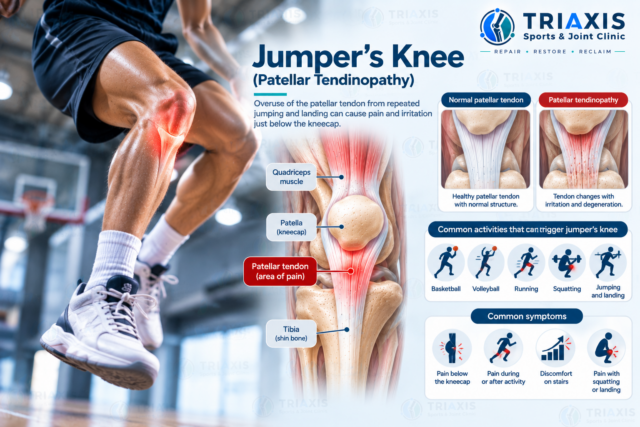
Jumper’s knee is the common name for patellar tendinopathy, a condition involving pain and changes in the patellar tendon. This tendon connects the kneecap to the shin bone and helps transfer force when you straighten your knee, run, jump and land. Symptoms often develop when repeated tendon loading exceeds its current capacity.
The patellar tendon runs from the patella (kneecap) to the upper part of the tibia (shin bone).
Together with the quadriceps muscles, quadriceps tendon and kneecap, it forms part of the knee’s extensor mechanism. This system allows you to straighten your knee and generate force during activities such as running, jumping, climbing stairs and getting up from a chair.
Jumper’s knee is one specific cause of knee pain. It should not be confused with runner’s knee, meniscus injuries or ligament injuries.
What does jumper’s knee feel like?
Jumper’s knee usually causes pain at the front of the knee, most commonly just below the kneecap where the patellar tendon begins. Pain may initially occur during or after demanding exercise and can become more noticeable with jumping, landing, running, squatting or other activities that load the tendon.
Common symptoms may include:
- Pain just below the kneecap
- Tenderness over the patellar tendon
- Pain during jumping or landing
- Pain during or after running
- Discomfort with squats or lunges
- Pain during stairs
- Reduced tolerance for sporting activity
Symptoms often develop gradually rather than after one clear injury.
Other causes of anterior knee pain include patellofemoral pain, fat pad problems and, less commonly, an acute tendon injury. Therefore, pain location should be interpreted together with history and examination.
What causes patellar tendinopathy?
Patellar tendinopathy is commonly associated with repeated high loads through the tendon, particularly jumping and landing. Changes in training volume, intensity or frequency may contribute when the tendon has insufficient time to adapt. However, jumper’s knee usually involves several interacting factors rather than one single cause.
Training-related factors may include:
- Increasing jumping volume
- More frequent training
- Increasing running or sprinting
- Introducing more explosive exercises
- Returning quickly after time away from sport
- Changing training surfaces or demands
Strength, jumping and landing mechanics, previous symptoms and recovery between training sessions may also be relevant.
Importantly, patellar tendinopathy should not simply be described as “inflammation”. Tendinopathy can involve changes in tendon structure and its response to repeated loading.
Which sports are commonly associated with jumper’s knee?
Jumper’s knee is particularly associated with sports involving repeated jumping, landing and explosive movements. Basketball and volleyball are common examples, although patellar tendon symptoms can also develop in football, badminton, tennis, running and other activities that repeatedly load the knee extensor mechanism.
Relevant sports may include:
- Basketball
- Volleyball
- Badminton
- Football and futsal
- Tennis
- Pickleball
- Padel
- Athletics
- Running
- Rugby
- Weightlifting
The sport itself does not mean someone will develop patellar tendinopathy. Training exposure, load changes, physical capacity and individual factors can all influence symptoms.
This is also why jumper’s knee can occur in active people who do not participate in traditional jumping sports.
Can jumper’s knee improve without surgery?
Yes. Jumper’s knee is managed without surgery. Treatment focuses on understanding and adjusting tendon load, followed by progressive strengthening to build the tendon’s capacity for everyday activity and sport, usually with a physiotherapist. Complete rest is not required, although temporarily reducing aggravating activities helps. Where tendon pain persists despite good rehabilitation, ultrasound-guided treatments can be added to support recovery.
Rehabilitation may include:
- Education about tendon loading
- Temporary modification of jumping or running
- Quadriceps strengthening
- Progressive tendon-loading exercises
- Hip and lower-limb strengthening
- Gradual reintroduction of jumping
- Landing and movement training
- Progressive return to sport
- Ultrasound-guided treatments where tendon pain persists despite rehabilitation
Exercise should be progressed according to symptoms, strength and sporting demands.
Some programmes use isometric, eccentric or heavy slow resistance exercises at different stages, and the programme is tailored to each person. A large living review of exercise treatments for lower-limb tendinopathy supports exercise as the first-line treatment, with additional treatments considered when progress is slow after around three months.
Can I continue playing sport with jumper’s knee?
Some people can continue modified training while managing jumper’s knee, while others may need to temporarily reduce activities that repeatedly aggravate the tendon. The decision depends on pain, tendon irritability, sporting demands and how symptoms respond during and after training. Complete withdrawal from all exercise is not always necessary.
Load modification could involve temporarily reducing:
- Number of jumps
- High-intensity training sessions
- Sprinting
- Plyometric exercises
- Deep or heavily loaded squats
- Consecutive training days
As tendon capacity improves, these activities can be progressively reintroduced.
Return to unrestricted sport should consider strength, jumping tolerance, landing control and the ability to complete sport-specific training without an unacceptable increase in symptoms.
How long does jumper’s knee take to improve?
Patellar tendinopathy can take time to improve because tendon adaptation occurs gradually. Recovery varies according to how long symptoms have been present, the level of sport, training demands, tendon capacity and consistency with rehabilitation. Rather than using one fixed timeline, progress is usually assessed through symptoms, strength and tolerance for increasingly demanding activity.
Signs of progress may include:
- Better tolerance for squatting
- Improved strength
- Less discomfort during everyday activities
- Increased running tolerance
- Ability to perform controlled jumping
- Progression to repeated jumping and landing
- Return to sport-specific training
Persistent tendinopathy can sometimes require several months of progressive rehabilitation.
Can ultrasound diagnose jumper’s knee, and when is MRI needed?
Jumper’s knee is diagnosed through a careful history and examination, and ultrasound is particularly well suited to examining the patellar tendon. Ultrasound may be useful, alongside clinical assessment, to confirm tendon changes, check for other causes of pain and guide treatment, often at the same visit. MRI is valuable when the diagnosis is uncertain or another knee condition is suspected. Imaging is always interpreted together with symptoms and examination.
An X-ray does not show patellar tendinopathy directly but may help assess bone and other joint-related conditions.
An MRI can provide detailed images of the patellar tendon and surrounding knee structures.
Musculoskeletal ultrasound provides real-time, high-resolution images of the patellar tendon. Radiology reviews describe ultrasound as particularly well suited to evaluating the patellar and quadriceps tendons [1]. It can show tendon thickening, changes in tendon structure, small tears and neovascularity (new blood vessel growth often seen in painful tendons), and can be repeated to monitor how the tendon responds to treatment. If a procedure is recommended, the same ultrasound is used to guide the needle precisely.
| Test | What it shows | When it may be useful |
| Ultrasound | Patellar tendon thickness and structure, neovascularity, small tears, nearby bursae and fat pad | Alongside examination to confirm tendon changes, monitor progress and guide procedures |
| MRI | The tendon plus cartilage, menisci, ligaments and bone | Uncertain diagnosis or suspected additional knee condition |
| X-ray | Bones only; the tendon is not visible | Suspected bony problem |
What non-surgical treatments can help jumper’s knee?
Progressive tendon-loading rehabilitation is the foundation of treatment. When tendon pain persists despite a good programme, or limits the ability to train, several ultrasound-guided treatments can be added to reduce pain, stimulate tendon healing and help athletes return to sport. At Triaxis, these are performed under real-time ultrasound so treatment reaches the affected part of the tendon precisely.
Platelet-rich plasma (PRP)
PRP is prepared from a small sample of your own blood and concentrated to contain growth factors involved in tendon repair, then injected into the affected part of the tendon under ultrasound guidance. A network meta-analysis of 19 randomised trials found that leukocyte-poor PRP combined with a high-volume injection improved tendon function more than non-injection treatment at six months [3]. In a randomised trial comparing PRP, needle tenotomy and a sham injection, clinical improvement was greatest with PRP [4].
Shockwave therapy
Extracorporeal shockwave therapy delivers pressure waves through the skin to the painful tendon, with no needles or downtime. A 2026 meta-analysis of 13 studies found that shockwave reduced average pain scores from 5.2 to 2.7 out of 10 and improved tendon function, both by more than the threshold patients notice as meaningful. Combining PRP with shockwave has been shown to relieve pain faster than PRP alone.
Prolotherapy (dextrose injections)
Prolotherapy uses a dextrose (sugar) solution injected into the affected area of the tendon to stimulate a healing response. In a study of 47 people with persistent patellar tendinopathy, ultrasound-guided dextrose injections roughly halved pain during sport, from 78 to 39 out of 100, and the tendon’s appearance on ultrasound improved, including reduced neovascularity.
High-volume injection
A high-volume injection places a larger volume of fluid alongside the tendon under ultrasound guidance, aiming to disrupt the painful new blood vessels and nerves that grow into a chronic tendon. It is often combined with PRP.
Tenex (ultrasonic tenotomy)
Tenex removes degenerate tendon tissue through a needle-sized incision using ultrasonic energy, under local anaesthetic and ultrasound guidance. In a series of high-level athletes with long-standing patellar tendinopathy, pain scores fell from 6.4 to 2.8 out of 10, and most returned to full sport after an average of about three and a half months.
Nerve hydrodissection
Small sensory nerves at the front of the knee, such as the infrapatellar branch of the saphenous nerve, can become irritated and add to pain below the kneecap. Ultrasound-guided hydrodissection uses fluid to free an irritated nerve from surrounding tissue.
| Treatment | What it involves | When it may be considered |
| Tendon-loading rehabilitation | Progressive isometric, heavy slow resistance and jumping exercises | Everyone with jumper’s knee; the foundation of recovery |
| PRP | Your own concentrated platelets injected into the tendon | Persistent tendon pain despite rehabilitation |
| Shockwave therapy | Non-invasive pressure waves applied to the tendon | Persistent pain; can be combined with PRP |
| Prolotherapy | Dextrose injected into the affected area of tendon | Persistent pain, particularly with neovascularity on ultrasound |
| High-volume injection | Larger volume of fluid alongside the tendon | Persistent pain with neovascularity; often with PRP |
| Tenex | Ultrasonic removal of degenerate tendon tissue | Long-standing tendon pain not settling with other treatments |
| Hydrodissection | Fluid used to free an irritated nerve | Nerve-related pain at the front of the knee |
All of these treatments work best alongside a continued tendon-loading programme, which is progressed afterwards to rebuild strength and jumping capacity.
When should I seek medical assessment for jumper’s knee?
Consider medical assessment when pain below the kneecap persists, repeatedly returns or begins limiting training, sport or everyday activities. Assessment may also help when the diagnosis is unclear or symptoms are not improving despite appropriate changes to activity and rehabilitation. Sudden severe pain after an injury requires a different assessment from gradual tendinopathy.
Seek assessment particularly if there is:
- Persistent or increasing tendon pain
- Difficulty progressing exercise
- Significant loss of strength
- Increasing swelling
- Difficulty walking
- A sudden pop or acute injury
- Inability to straighten the knee normally
- Symptoms that do not fit typical patellar tendinopathy
Sudden pain with immediate loss of function may indicate an acute tendon injury rather than jumper’s knee.
How’s jumper’s knee assessed at Triaxis Sports & Joint Clinic?
Assessment of jumper’s knee at Triaxis Sports & Joint Clinic focuses on confirming whether symptoms arise from the patellar tendon and understanding the activities that load it. Where helpful, diagnostic ultrasound of the tendon can be performed at the same visit, so findings and treatment options can be discussed straight away. Assessment also considers pain location, training history, strength, movement and the person’s sporting or occupational demands.
Depending on the presentation, this may include:
- Sports Medicine assessment
- Musculoskeletal examination
- Patellar tendon assessment
- Knee and hip movement
- Lower-limb strength
- Squat and single-leg function
- Jumping and landing demands
- Training-load history
- Same-visit diagnostic ultrasound of the patellar tendon where appropriate
- Rehabilitation planning, coordinated with physiotherapy
- Ultrasound-guided treatments where tendon pain persists
- Patient education
- Shared decision-making
Frequently Asked Questions About Jumper’s Knee
Is jumper’s knee the same as patellar tendinopathy?
Yes. Jumper’s knee is the commonly used term for patellar tendinopathy. The condition typically causes pain in the patellar tendon, particularly near its attachment just below the kneecap. The term “jumper’s knee” reflects its association with jumping sports, although people who do not regularly jump can also develop patellar tendon symptoms.
Is jumper’s knee the same as runner’s knee?
No. Jumper’s knee usually refers to patellar tendinopathy, which affects the tendon below the kneecap. Runner’s knee commonly refers to patellofemoral pain, which tends to cause pain around or behind the kneecap. Their symptoms and aggravating activities can overlap, so clinical assessment may help distinguish between them.
Can I run with jumper’s knee?
Some people can continue running with temporary adjustments if symptoms remain manageable. Others may need to reduce running volume, speed or frequency while gradually rebuilding tendon capacity. Running is only one source of patellar tendon load, so the overall programme should also consider jumping, gym training and other sporting activities.
Are squats good for jumper’s knee?
Squatting can form part of rehabilitation because it loads the quadriceps and patellar tendon. However, the appropriate depth, resistance and number of repetitions depend on symptoms and current capacity. Heavier or deeper squats may aggravate a sensitive tendon, so rehabilitation usually progresses loading rather than immediately avoiding or maximising it.
Should I completely rest jumper’s knee?
Complete rest is not automatically necessary. Temporarily reducing activities that repeatedly aggravate symptoms may help, while an appropriate exercise programme maintains and progressively develops strength. Long periods of complete inactivity can reduce physical capacity, so management generally aims to balance symptom control with appropriate tendon loading.
Does jumper’s knee show up on ultrasound?
Yes. The patellar tendon lies close to the skin, which makes it well suited to ultrasound [1]. Ultrasound can show tendon thickening, structural changes, small tears and neovascularity, and can be repeated to monitor how the tendon responds to treatment. As tendon changes can sometimes be present without pain, ultrasound findings are interpreted together with your history and examination.
Can PRP or other injections help jumper’s knee?
Yes, for persistent tendon pain. A network meta-analysis of randomised trials found that leukocyte-poor PRP, alone or combined with a high-volume injection, improved pain and function compared with non-injection treatment at six months [3], and ultrasound-guided dextrose injections have been associated with less pain and an improved tendon appearance on ultrasound [2]. Injections work best alongside a structured tendon-loading programme.
What if my jumper’s knee is not improving?
If tendon pain persists despite a well-structured programme, the first step is to review the diagnosis, training load and the exercise programme itself. Ultrasound can help, alongside examination, to assess the tendon. Treatments such as PRP, shockwave therapy, prolotherapy, high-volume injection or Tenex can then be added to reduce pain and help you return to sport.
About the Author
Dr Dinesh Sirisena is a Consultant in Sports, Exercise & Musculoskeletal Medicine. His clinical work includes the assessment and management of sports injuries and musculoskeletal conditions involving muscles, tendons, ligaments, joints and related structures.
Medical Disclaimer
This article provides general educational information and does not replace individual medical assessment, diagnosis or treatment. Pain at the front of the knee can have several possible causes. Decisions about exercise, imaging, rehabilitation, procedures or further management should depend on the individual’s symptoms, medical history and clinical findings.