
Meniscus Tear: Causes, Symptoms and Treatment Options
Written and medically reviewed by
Dr Dinesh Sirisena
Consultant in Sports, Exercise & Musculoskeletal Medicine
Triaxis Sports & Joint Clinic
A meniscus tear affects one of the two cartilage structures that help distribute load inside the knee. Tears can occur suddenly during twisting or sport or develop gradually with age-related changes. Symptoms may include knee pain, swelling, catching, stiffness or locking. Most meniscus tears can be managed non-surgically. Diagnostic ultrasound in clinic can assess the meniscus at the first visit, and exercise-based rehabilitation the first-line treatment. For tears that stable but painful, ultrasound-guided injections such as platelet-rich plasma (PRP), hyaluronic acid or corticosteroid offer an effective option to ease pain and support recovery.
What’s a meniscus tear?
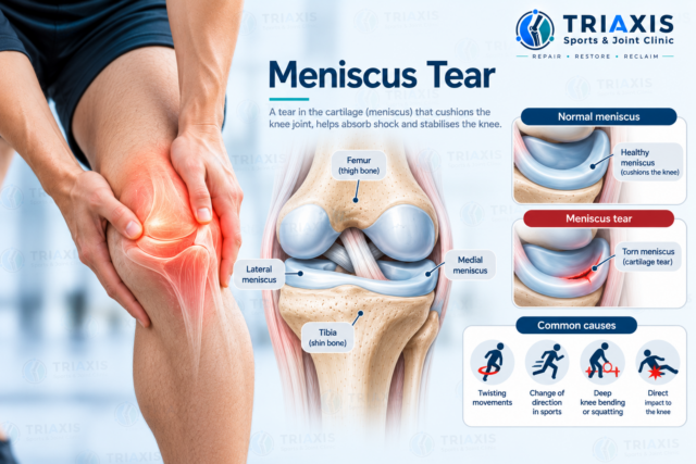
A meniscus tear an injury or structural change affecting one of the two menisci inside the knee. The menisci C-shaped pieces of fibrocartilage between the thigh bone and shin bone. They help distribute load, absorb forces and contribute to knee stability during walking, running and sport.
The knee contains a medial meniscus on the inner side and a lateral meniscus on the outer side.
They sit between the femur (thigh bone) and tibia (shin bone). Other important structures include the articular cartilage, ACL, PCL, MCL and LCL, together with the muscles and tendons surrounding the knee.
Because these structures work together, a meniscus injury can sometimes occur alongside a knee ligament injury. Triaxis also identifies meniscus injuries as one of several possible causes of knee pain.
What does a meniscus tear feel like?
A meniscus tear may cause pain along the inner or outer joint line of the knee, depending on which meniscus affected. Some people develop swelling, stiffness, catching or difficulty fully bending or straightening the knee. Symptoms vary considerably, and not every meniscus tear produces locking or severe pain.
Possible symptoms include:
- Inner or outer knee pain
- Swelling
- Stiffness
- Pain during twisting
- Pain with deep squatting
- Catching or clicking
- Difficulty fully bending or straightening the knee
- A sensation that something blocking movement
These symptoms not specific to the meniscus. Ligaments, cartilage, tendons and osteoarthritis can produce similar knee pain.
What causes a meniscus tear?
Meniscus tears may occur after a sudden twisting injury or develop gradually as the meniscus changes over time. Acute tears more commonly associated with sport or trauma, while degenerative tears more common with increasing age and may occur during relatively ordinary movements.
An acute tear may happen when the foot remains planted while the knee twists. Deep bending or forceful rotation can also place stress on the meniscus.
Sports that involve pivoting, cutting, landing or contact may be associated with meniscus injuries, including:
- Football and futsal
- Basketball
- Rugby
- Badminton
- Tennis
- Pickleball
- Padel
Weightlifting movements involving deep knee flexion may aggravate an already symptomatic meniscus in some people, although squatting itself does not mean someone will develop a tear.

Meniscus injuries can also occur alongside ACL and other sports-related knee injuries.
Can a meniscus tear improve without surgery?
Most meniscus tears can be managed without surgery, particularly degenerative tears and acute tears that are stable, meaning the torn part is not displaced and the knee is not locking.
Management includes education, temporary activity modification and progressive exercise-based rehabilitation. For stable tears that remain painful, ultrasound-guided injections can be added to ease pain and help rehabilitation progress. The decision depends on the type of tear, symptoms, knee function and associated injuries.
Rehabilitation may focus on:
- Restoring comfortable knee movement
- Managing swelling
- Quadriceps strengthening
- Hamstring strengthening
- Hip and gluteal strength
- Balance and single-leg control
- Gradual return to running
- Progressive sport-specific movements
- Ultrasound-guided injections for stable but painful tears
For degenerative meniscus problems, an international consensus of 67 experts from 14 countries recommends exercise-based rehabilitation as the first-line approach, and notes it may also be an option for some acute tears [1].
Further assessment or management may be needed where symptoms and clinical findings suggest a mechanically unstable tear, such as one that locks the knee.
How long does a meniscus tear take to recover?
There’s no single recovery time for every meniscus tear. Recovery depends on the tear type and location, whether it occurred suddenly or gradually, associated knee injuries, symptoms and activity demands. People treated with rehabilitation, with or without injections, progress according to function rather than a fixed timeline.
A small stable tear and a displaced traumatic tear are very different injuries.
Return to activity should therefore consider:
- Pain and swelling
- Knee movement
- Strength
- Walking and running ability
- Balance
- Change-of-direction control
- Sport-specific function
A reduction in pain alone does not necessarily mean the knee is ready for unrestricted sport.
Can ultrasound diagnose a meniscus tear, and when is MRI needed?
Yes, in experienced hands. High-resolution musculoskeletal ultrasound may be useful in detecting meniscus tears, along with clinical assessment, and in assessing the structures around them. Often at the same visit as the clinical examination.
MRI remains valuable for mapping complex or displaced tears and for checking the ACL, cartilage and bone. Imaging is always interpreted together with symptoms and examination.
An X-ray does not directly show a meniscus tear. It may be considered to assess bones, joint alignment or osteoarthritis.
An MRI can visualise the medial and lateral menisci as well as cartilage, ligaments and other structures inside the knee.
Musculoskeletal ultrasound provides real-time images of the meniscus edges and the joint line. A 2025 meta-analysis of 11 studies and 866 knees found ultrasound performed by experienced operators had a pooled accuracy of about 95% for meniscus tears, with no significant difference in sensitivity or specificity compared with MRI.
Ultrasound also shows features that help guide treatment. It can measure meniscal extrusion, where the meniscus is pushed out from the joint and which may point to a root tear, identify parameniscal cysts, assess the joint for swelling and examine nearby ligaments and tendons that may be causing similar pain [3]. If an injection is recommended, the same ultrasound is used to guide the needle precisely to the meniscus or joint.
| Test | What it shows | When it is most useful |
| Ultrasound | Meniscus tears near the joint line, extrusion, parameniscal cysts, swelling, nearby ligaments and tendons | First-line assessment at the clinic visit; guiding injections |
| MRI | The full meniscus, including deeper and complex tears, plus ACL, cartilage and bone | Locking, suspected displaced or root tears, or associated ligament injury |
| X-ray | Bones, alignment and osteoarthritis; the meniscus is not visible | Assessing arthritis, especially in older adults |
Importantly, an MRI finding should be interpreted together with the person’s symptoms and examination. Meniscal changes in imaging do not always explain the person’s pain.
What treatment options are available for a meniscus tear?
Treatment depends on whether the tear is acute or degenerative, its location and pattern, the person’s symptoms and whether other knee structures are injured. A useful starting point is whether the tear is stable or mechanically unstable. Stable tears, which make up the majority, are managed with rehabilitation and, where pain persists, ultrasound-guided injections. Tears that lock or block the knee may need further assessment or management.
Management includes education, modifying aggravating activities, progressive rehabilitation and, for stable but painful tears, ultrasound-guided injections.
Further assessment or management may be needed if there is:
- Persistent mechanical restriction or locking
- A displaced tear
- Significant acute trauma
- An associated ligament injury
The aim is not simply to treat an MRI image. Management reflects the person’s symptoms, knee function and goals.
| Stable but painful tear | Mechanically unstable tear | |
| Typical features | Joint-line pain, swelling or catching; knee still moves fully | True locking, a block to straightening, or a displaced fragment |
| Common examples | Degenerative and horizontal tears; small or partial acute tears | Bucket-handle and displaced flap tears |
| Usual first approach | Rehabilitation, with ultrasound-guided injections if pain persists | Further assessment or management may be needed |
Can injections help a stable but painful meniscus tear?
Yes. For stable meniscus tears that remain painful despite rehabilitation, or that are too painful to rehabilitate effectively, ultrasound-guided injections are an effective option. They aim to reduce pain and inflammation, support the healing environment of the knee and allow strengthening to progress, helping people return to the activities they enjoy.
A tear considered stable when the torn part not displaced and the knee not locking or blocked. Ultrasound and clinical examination help confirm this before any injection. At Triaxis, all knee injections are performed under ultrasound guidance; research has shown that ultrasound can accurately and safely guide a needle into the body and back of both menisci [7].
Platelet-rich plasma (PRP)
PRP is prepared from a small sample of your own blood and concentrated to contain growth factors involved in tissue repair. It can be injected into the knee joint, directly into the torn meniscus under ultrasound guidance, or both. A 2024 systematic review of 10 studies and 686 patients found that most studies showed improved pain and function by three months, lasting at least one year [4]. In a series of 392 patients treated with combined intrameniscal and intra-articular PRP, 90% continued to be managed without further intervention at follow-up, with the best results seen in horizontal tears. A randomised controlled trial found that adding PRP to a needling procedure improved healing of chronic meniscus tears, with fewer patients needing further intervention (8% versus 28%).
Hyaluronic acid (HA)
Hyaluronic acid is a lubricating, cushioning substance naturally found in joint fluid. An intra-articular HA injection can improve joint lubrication and ease pain, and particularly useful for degenerative meniscus tears where early wear of the joint surface also present. Recent expert reviews describe injections such as hyaluronic acid and corticosteroid as effective first-line options for atraumatic (degenerative) meniscus tears.
Corticosteroid injection
A corticosteroid injection reduces inflammation and can settle a painful flare quickly, creating a window in which rehabilitation can progress. Ultrasound-guided injections around the meniscus have been reported to provide short-term relief of meniscus symptoms [3]. Corticosteroid used selectively, as it acts mainly on pain and inflammation rather than tissue healing.
Prolotherapy
Prolotherapy uses a dextrose (sugar) solution to stimulate a local healing response. It may be considered when the small ligaments that anchor the meniscus to the shin bone, or the joint capsule, contribute to persistent pain. A 2026 case report described improvements in the meniscus, surrounding knee structures and function after a course of ultrasound-guided dextrose injections combined with exercise.
Nerve hydrodissection
Some people with long-standing inner knee pain develop irritation of small sensory nerves around the knee, such as the infrapatellar branch of the saphenous nerve. Ultrasound-guided hydrodissection uses fluid to free an irritated nerve and may help where nerve-type pain continues after the meniscus has settled.
| Treatment | What it involves | When it may be considered |
| Rehabilitation | Progressive exercise programme with a physiotherapist | All meniscus tears; the foundation of recovery |
| PRP | Your own concentrated platelets injected into the joint and/or the torn meniscus | Stable tears with persistent pain, especially degenerative and horizontal tears |
| Hyaluronic acid | Lubricating injection into the joint | Degenerative tears, particularly with early joint wear |
| Corticosteroid | Anti-inflammatory injection into or around the joint | A painful flare limiting rehabilitation |
| Prolotherapy | Dextrose injections around supporting ligaments or capsule | Persistent pain from the meniscal ligaments or capsule |
| Hydrodissection | Fluid used to free an irritated nerve | Nerve-type knee pain that persists |
Injections are best suited to stable tears. If the knee locks or a tear appears displaced, further assessment or management may be needed.
When should I seek medical assessment for a possible meniscus tear?
Consider medical assessment when knee pain or swelling persists after a twisting injury, movement remains restricted, or symptoms interfere with walking, exercise or sport. Assessment particularly important if the knee becomes mechanically locked, significant swelling follows trauma or you cannot comfortably bear weight.
Assessment may also be appropriate for:
- Repeated catching or locking
- Persistent joint-line pain
- Recurrent swelling
- Difficulty fully straightening the knee
- Knee instability
- Symptoms that do not improve with activity modification
- A significant sporting injury
A locked knee after an acute injury warrants timely assessment because certain displaced tears may physically restrict movement.
How’s a meniscus tear assessed at Triaxis Sports & Joint Clinic?
Assessment at Triaxis Sports & Joint Clinic focuses on determining whether symptoms arise from the meniscus or another knee structure, and whether a tear stable. Diagnostic ultrasound usually performed at the same visit, so findings and treatment options can be discussed straight away. Clinical assessment also considers the injury mechanism, pain pattern, swelling, knee movement, strength, stability and the demands of the person’s work, exercise or sport.
Depending on the findings, assessment may include:
- Sports Medicine assessment
- Musculoskeletal examination
- Knee movement and joint-line assessment
- Ligament stability testing
- Strength and functional assessment
- Review of sporting or activity demands
- Review of existing imaging
- Same-visit diagnostic ultrasound of the meniscus and surrounding structures
- Rehabilitation planning, coordinated with physiotherapy
- Ultrasound-guided injections for stable but painful tears where appropriate
- MRI referral where a displaced tear or ligament injury suspected
- Patient education
- Shared decision-making
Further assessment or management discussed where the clinical findings suggest a mechanically unstable tear.
Frequently Asked Questions About Meniscus Tears
Can I walk with a meniscus tear?
Some people can walk relatively normally with a meniscus tear, while others experience pain, swelling or restricted movement. Walking ability does not determine the severity of a tear by itself. If weight-bearing difficult after an acute injury, the knee repeatedly catches, or movement significantly restricted, medical assessment may be appropriate.
Where does a meniscus tear usually hurt?
A medial meniscus injury may cause pain around the inner joint line, while a lateral meniscus injury may cause pain along the outer joint line. However, pain location alone cannot be diagnosed with a meniscus tear because ligaments, tendons, cartilage and other knee structures may cause symptoms in similar areas.
Can a meniscus tear heal by itself?
The potential for biological healing depends partly on the tear’s location and blood supply. The outer part of the meniscus has a better blood supply than the inner region. Importantly, symptom improvement does not require the meniscus to return to its previous appearance on imaging, and many people regain good function with rehabilitation. For stable tears, PRP being used to support the healing environment, with studies reporting improved pain and function [4, 5].
Is a meniscus tear always painful?
No. Meniscal changes can sometimes be present on MRI without causing symptoms. This particularly relevant with age-related or degenerative changes. For this reason, clinicians generally interpret MRI findings together with symptoms, examination findings and function rather than assuming that every meniscus tear seen on a scan is the source of pain.
Should I avoid squats with a meniscus tear?
Not necessarily. Deep knee bending may aggravate symptoms in some people, particularly during the more sensitive stages of an injury. Rehabilitation can modify depth, load or exercise selection before gradually progressing according to symptoms and function. Completely avoiding squatting long-term is not automatically necessary for every meniscus tear.
Can I run with a meniscus tear?
Whether running is appropriate depends on symptoms, swelling, strength and knee function. Running may temporarily need to be reduced if it repeatedly aggravates the knee. Rehabilitation can gradually progress from strengthening and lower-impact activity towards jogging and running when appropriate, followed by change-of-direction work for athletes.
What treatment does a meniscus tear need?
Most degenerative tears and many acute tears are managed with exercise-based rehabilitation. For stable tears that remain painful, ultrasound-guided injections such as PRP, hyaluronic acid or corticosteroid offer a further step to ease pain and support recovery. Tears that lock or block the knee may need further assessment or management.
What’s the difference between a meniscus tear and an ACL injury?
The meniscus fibrocartilage that helps distribute forces inside the knee, while the ACL a ligament that contributes to knee stability. A meniscus tear may cause joint-line pain, swelling or catching, whereas an ACL injury commonly causes instability after a pivoting injury. Both structures can be injured during the same event.
Can ultrasound detect a meniscus tear?
Yes. When performed by an experienced clinician, ultrasound detects meniscus tears with accuracy comparable to MRI; a 2025 meta-analysis reported a pooled accuracy of about 95% [2]. Ultrasound can also show meniscal extrusion, cysts and swelling, and used to guide injections. MRI remains useful for complex, displaced or deeper tears and for checking the ACL and cartilage.
Can injections help a stable meniscus tear?
Yes. For stable tears that remain painful, ultrasound-guided injections are an established option. PRP has been associated with improved pain and function and fewer people needing further intervention [4, 5, 6], while hyaluronic acid and corticosteroid can ease pain and help rehabilitation progress [8]. Injections work best alongside a structured exercise programme.
About the Author
Dr Dinesh Sirisena a Consultant in Sports, Exercise & Musculoskeletal Medicine. His clinical work includes the assessment and management of sports injuries and musculoskeletal conditions involving joints, muscles, tendons, ligaments and related structures.
Medical Disclaimer
This article provides general educational information and does not replace individual medical assessment, diagnosis or treatment. Knee symptoms can arise from several structures and conditions. Decisions about imaging, rehabilitation, procedures or further management should depend on the individual’s symptoms, medical history, examination findings and activity requirements.