
Medial Collateral Ligament (MCL) Injury: Causes, Symptoms and Treatment Options
Written and medically reviewed by Dr Dinesh Sirisena
Medial collateral ligament (MCL) injury
Consultant in Sports, Exercise & Musculoskeletal Medicine
Triaxis Sports & Joint Clinic
A medial collateral ligament (MCL) injury affects the ligament on the inner side of the knee. It commonly causes inner knee pain, tenderness, swelling and sometimes a feeling of instability. MCL injuries range from mild sprains to complete tears. Most isolated MCL injuries heal well without surgery. Diagnostic ultrasound in clinic can show the injured ligament in real time, often at the first visit, and a structured rehabilitation programme forms the foundation of recovery. For injuries that are slow to settle, ultrasound-guided platelet-rich plasma (PRP) or prolotherapy may be added to support ligament healing and a return to sport.
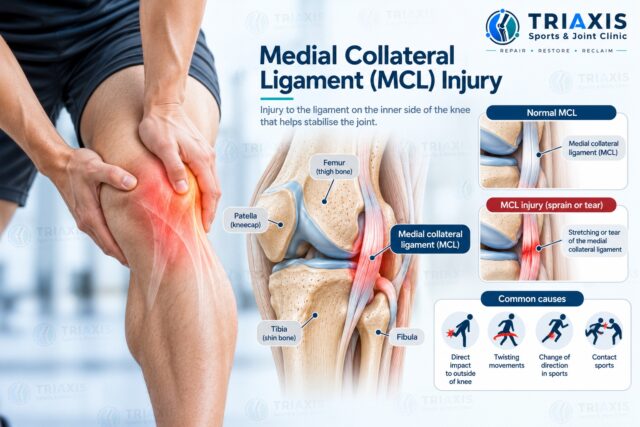
What’s the medial collateral ligament and what happens when it is injured?
The medial collateral ligament, or MCL, a band of tissue that helps stabilise the inner side of the knee. An MCL injury occurs when the ligament stretched or torn.
Injury may range from a mild sprain to a complete tear, depending on the force involved and the extent of ligament damage.
MCL connects the femur (thigh bone) to the tibia (shin bone) along the inner side of the knee. It helps resist forces that push the knee inward.
It works with the other major knee ligaments — the ACL, PCL and LCL — to maintain knee stability. You can learn more about how these structures differ in our guide to knee ligament injuries.
MCL injuries are commonly classified as:
Firstly – Grade 1: The ligament mildly sprained but remains stable.
Secondly – Grade 2: The ligament partially torn and may have some looseness.
Lastly – Grade 3: The ligament completely torn and may result in greater instability.
The injury grade only one part of the assessment. Symptoms, knee stability, other injured structures and activity requirements also matter.
What does an MCL injury feel like?
An MCL injury usually causes pain and tenderness along the inner side of the knee.
Swelling and stiffness may develop after the injury. With a more significant tear, the knee may feel unstable or give way, particularly during turning, sideways movements or changes of direction.
Common symptoms include:
- Inner knee pain
- Tenderness over the MCL
- Local swelling
- Knee stiffness
- Pain when walking
- Pain when bending the knee
- Difficulty changing direction
- A feeling of instability or giving way
However, not every case of inner knee pain caused by MCL. The medial meniscus, tendons and joint surfaces can produce symptoms in a similar area.
What causes an MCL injury?
An MCL injury commonly occurs when a force pushes the knee inward, placing stress on the ligament along the inner knee.
A direct impact on the outside of the knee is one common mechanism. MCL injuries may also occur during twisting, awkward landings or rapid changes of direction.
This explains why MCL injuries are often associated with contact and change-of-direction sports.
Possible mechanisms include:
- A collision affecting the outside of the knee
- Sudden twisting
- Pivoting
- Awkward landing
- Rapid change of direction
- Falls or other trauma
An MCL injury may occur on its own or together with an ACL, meniscus or other knee injury.
Which sports are associated with MCL injuries?
Sports involving physical contact, pivoting, cutting or sudden changes of direction can place greater stress on the MCL. Football, futsal, rugby and basketball are common examples, although MCL injuries can also occur in racquet sports, skiing and other activities involving unexpected sideways knee forces.
Following, relevant activities may include:
- Football and futsal
- Rugby
- Basketball
- Badminton
- Tennis
- Pickleball
- Padel
- Skiing
- Martial arts
Running in a straight line generally places different demands on the knee, although an MCL injury can still occur during running if there’s a fall, collision or sudden change of direction.
The broader Triaxis sports medicine and injury services page explains how different sporting and musculoskeletal injuries may be assessed.
Can an MCL injury improve without surgery?
Most isolated MCL injuries heal well without surgery. Treatment focuses on protecting the knee when necessary, restoring movement and progressively rebuilding strength and stability, usually with a physiotherapist or other rehabilitation professional.
A brace may be used for some injuries. Where recovery stalls, ultrasound-guided PRP or prolotherapy can be added to the rehabilitation plan. Surgery reserved for selected cases, such as certain severe or combined ligament injuries.
Following, management may include:
- Education about the injury
- Temporary activity modification
- Swelling management
- Progressive weight-bearing as appropriate
- Restoring knee movement
- Quadriceps and hamstring strengthening
- Hip and lower-limb strengthening
- Balance and coordination exercises
- Bracing where appropriate
- Gradual return to running and sport
- Ultrasound review to monitor ligament healing
- Ultrasound-guided PRP or prolotherapy where recovery slower than expected
An isolated MCL injury different from a knee injury involving several ligaments. Combined injuries require individual assessment because management may change.
How long does an MCL injury take to recover?
Recovery depends on the severity and location of the MCL injury, whether other knee structures are involved and the person’s physical demands. A mild sprain generally recovers sooner than a complete tear. Return to sport should consider knee function, strength and stability rather than relying only on a fixed number of weeks.
Before progressing back to demanding sport, rehabilitation may assess:
- Pain and swelling
- Knee range of motion
- Muscle strength
- Knee stability
- Balance and single-leg control
- Running ability
- Jumping and landing
- Change-of-direction ability
- Sport-specific movements
Feeling less pain does not necessarily mean the ligament and knee are ready for unrestricted sport.
Can ultrasound diagnose an MCL injury, and when MRI needed?
Yes. Because the MCL lies close to the skin, high-resolution musculoskeletal ultrasound well suited to examining it.
Ultrasound can show whether the ligament thickened, partially torn or completely torn, and can assess it while the knee gently stressed. It’s quick, radiation-free and can often be done at the same visit as the clinical examination.
MRI valuable when a deeper injury, such as an ACL, meniscus or cartilage injury, suspected.
An X-ray does not show the MCL directly. However, it may be useful when a fracture or other bone injury suspected.
An MRI provides detailed images of the MCL and deeper structures such as the ACL, PCL, menisci and cartilage.
Musculoskeletal ultrasound provides real-time, high-resolution images of the MCL. In a study of 250 patients with acute knee trauma, ultrasound identified MCL tears with an overall accuracy of 92% compared with MRI, and a normal scan correctly ruled out an MCL tear in 95% of cases [2]. Radiology reviews describe ultrasound as particularly well suited to the medial and lateral collateral ligaments.
Ultrasound also offers practical advantages that MRI cannot. It’s dynamic: the ligament can be watched while a gentle sideways (valgus) stress applied, and the injured knee can be compared directly with the other side. It can be repeated during recovery to track ligament healing and help guide rehabilitation progression [4]. And if a procedure recommended, the same ultrasound can be used to guide the needle precisely to the injured area.
| Test | What it shows | When it’s most useful |
| Ultrasound | MCL thickening, partial or complete tears, stress gapping, swelling, nearby tendons and bursae | First-line imaging of the MCL; monitoring healing; guiding procedures |
| MRI | MCL plus deeper structures: ACL, PCL, menisci, cartilage and bone bruising | Suspected combined injury, marked instability or unclear diagnosis |
| X-ray | Bones only; the MCL itself not visible | Suspected fracture or bony injury after significant trauma |
What exercises may help after an MCL injury?
Rehabilitation the foundation of MCL recovery.
A programme, usually delivered with a physiotherapist, progresses from restoring comfortable knee movement and muscle activation towards strengthening, balance and more demanding functional exercises. Later stages include running, jumping and change-of-direction training.
The programme reflects the severity of the injury and the person’s activity goals, and continues alongside any injection treatment.
Generally exercises may gradually address:
- Knee movement
- Quadriceps strength
- Hamstring strength
- Hip and gluteal strength
- Calf strength
- Balance
- Single-leg control
- Running mechanics
- Jumping and landing
- Change of direction
Meanwhile, an athlete returning to football or rugby will usually have different functional requirements from someone whose main goal comfortable walking and everyday activity.

What non-surgical treatments & procedures can help an MCL injury?
Rehabilitation remains the cornerstone of MCL care, and most people recover well with it.
When pain, tenderness or looseness persists, or an athlete needs to return to sport, several ultrasound-guided treatments can be added to support ligament healing.
Triaxis Treatments Options: These delivered under real-time ultrasound so the treatment reaches the injured part of the ligament precisely.
Platelet-rich plasma (PRP)
PRP prepared from a small sample of your own blood, concentrated to contain growth factors involved in tissue repair. It’s injected under ultrasound guidance directly into the injured area of the MCL.
A 2025 report from a Singapore university hospital described two patients with persistent deep MCL injuries, a young footballer and an older walker, who both had marked improvements in pain and function within four weeks of ultrasound-guided PRP; the footballer returned to his sport. Reviews of PRP in ligament injuries report a quicker return to daily activities with a favourable safety profile. PRP typically considered for partial tears or chronic MCL pain that has not settled with rehabilitation.
Prolotherapy
Prolotherapy involves injecting a dextrose (sugar) solution around the injured ligament to stimulate a local healing response. Forthwith, it has a long history in ligament and joint-stability problems. In laboratory studies, dextrose injections increased the thickness of injured MCLs, and a published case of a rugby player with a grade 2 MCL sprain described a pain-free return to full rugby movements after three weeks of prolotherapy combined with exercise therapy. Prolotherapy may be considered for persistent MCL pain or a sense of looseness, often as a course of injections.
Ultrasound-guided injections
Therefore, some people with an MCL injury develop irritation of nearby structures, such as the bursa beneath the MCL or the pes anserine bursa just below it. A targeted ultrasound-guided injection can settle this irritation so that rehabilitation can progress comfortably. Corticosteroid not injected into the ligament itself.
Shockwave therapy
Extracorporeal shockwave therapy delivers pressure waves through the skin to areas of long-standing pain. It may be considered for chronic pain where the MCL attaches to the thigh bone, and requires no needles or downtime.
Nerve hydrodissection
A small sensory branch of the saphenous nerve runs close to the inner knee and can become irritated or trapped by scar tissue after injury, causing burning or sharp inner knee pain. Ultrasound-guided hydrodissection uses fluid to gently free the nerve from surrounding tissue.
Tenex (ultrasonic tenotomy)
Tenex treats the tendon rather than the ligament. It may be considered when a nearby tendon, such as the semimembranosus or pes anserine tendons, develops long-standing tendinopathy alongside or after an MCL injury. Tenex therapy removes degenerate tendon tissue through a small needle-sized incision under ultrasound guidance.
| Treatment | What it involves | When it may be considered |
| Rehabilitation | Progressive exercise programme with a physiotherapist | All MCL injuries; the foundation of recovery |
| PRP | Your own concentrated platelets injected into the MCL under ultrasound | Partial tears or chronic MCL pain not settling with rehabilitation |
| Prolotherapy | Dextrose injections around the ligament | Persistent MCL pain or a feeling of looseness |
| Ultrasound-guided injection | Targeted injection of an irritated bursa | Bursitis limiting rehabilitation |
| Shockwave therapy | Non-invasive pressure waves | Chronic pain at the MCL attachment |
| Hydrodissection | Fluid used to free an irritated nerve | Nerve-type inner knee pain after injury |
| Tenex | Removal of degenerate tendon tissue | Associated chronic tendinopathy near the MCL |
Surgery reserved for selected severe, unstable or combined ligament injuries, and a surgical opinion arranged where appropriate.
When should I seek medical assessment for an MCL injury?
Medical assessment may be appropriate when inner knee pain, swelling or instability persists after an injury, particularly if walking or normal activity difficult. Assessment also useful when the knee repeatedly gives way or symptoms suggest that the ACL, meniscus or another structure may have been injured at the same time.
Following assessment if there is:
- Significant or persistent inner knee pain
- Difficulty bearing weight
- Marked swelling
- Knee instability or giving way
- Reduced movement
- Locking or catching
- A pop or tearing sensation during injury
- Symptoms that are not improving
More urgent assessment may be appropriate following major trauma, particularly if the knee appears deformed or weight-bearing not possible.
How’s an MCL injury assessed at Triaxis Sports & Joint Clinic?
Assessment of a suspected MCL injury at Triaxis Sports & Joint Clinic focuses on identifying the injured structures, examining knee stability and understanding how the injury affects everyday activity or sport. Diagnostic ultrasound is usually performed at the same visit, so the findings and the treatment plan can be discussed straight away. Because MCL injuries may occur together with other knee injuries, the assessment considers the knee as a whole.
Depending on the clinical situation, this may include:
- Sports Medicine assessment
- Musculoskeletal examination
- Knee movement and swelling assessment
- Ligament stability testing
- Strength and functional assessment
- Review of the injury mechanism
- Same-visit diagnostic ultrasound, including dynamic stress imaging
- MRI referral where a combined injury suspected
- Rehabilitation planning, coordinated with physiotherapy
- Ultrasound-guided PRP, prolotherapy or other procedures where appropriate
- Follow-up ultrasound to monitor healing
- Patient education
- Shared decision-making
Surgical assessment arranged where appropriate, particularly for selected severe, persistent or combined ligament injuries.
Frequently Asked Questions About MCL Injuries
Can I walk with an MCL injury?
Some people with a mild MCL injury can walk, although the knee may feel sore or stiff. More significant injuries can make weight-bearing uncomfortable or cause instability. Temporary activity modification or bracing may be considered in some cases. If walking very difficult after an injury, or the knee repeatedly gives way, clinical assessment may help determine the extent of the injury.
Where does an MCL tear hurt?
An MCL injury usually causes pain and tenderness along the inner side of the knee. The exact location may vary depending on which part of the ligament affected. However, the medial meniscus, tendons and other structures can also cause inner knee pain. Pain location alone therefore cannot confirm that the MCL injured.
Is an MCL tear worse than an ACL tear?
Both MCL and ACL injuries affect different structures and should not simply be classified as one being worse than the other. Many isolated MCL injuries can be managed without surgery. ACL injuries may cause a different pattern of instability and may require different management depending on activity demands. When both ligaments are injured, the treatment plan may change.
Does a Grade 3 MCL tear always require surgery?
No. A Grade 3 injury describes a complete MCL tear, but it does not automatically mean surgery required. Some isolated complete tears may be managed without an operation. The tear location, knee stability, other ligament injuries, activity demands and response to rehabilitation may all influence whether surgical assessment appropriate.
Should I wear a knee brace for an MCL injury?
A knee brace may be considered for selected MCL injuries to help limit unwanted sideways stress while allowing appropriate movement. However, bracing requirements differ according to injury severity and clinical findings. There’s no single bracing programme that’s appropriate for every MCL injury, so the decision should be individualised.
Can I run with an MCL injury?
Running may need to be temporarily reduced after an MCL injury, particularly if it causes pain, swelling or instability. A gradual return usually considers comfortable walking, knee movement, strength and stability before progressing to jogging and faster running. Athletes may require further change-of-direction and sport-specific rehabilitation before returning to unrestricted sport.
Can an MCL injury happen without contact?
Yes. Although a blow to the outside of the knee a common cause, MCL injuries can also occur without direct contact. Sudden twisting, awkward landings or rapid changes of direction may place enough stress on the inner knee to injure the ligament, particularly when the knee moves inward unexpectedly.
Would an MCL injury happen together with an ACL tear?
Possible. Both MCL and ACL can be injured during the same incident, and the meniscus or other knee structures may also be involved. Combined ligament injuries can affect knee stability and may require a different management plan from an isolated MCL injury. Clinical examination and, where appropriate, imaging can help determine which structures are affected.
Can ultrasound diagnose an MCL tear?
Yes. Evidently the MCL sits close to the skin, which makes it well suited to high-resolution ultrasound. Ultrasound can show the grade of injury, assess the ligament under gentle stress and compare it with the other knee. In a study of 250 patients with acute knee injuries, ultrasound identified MCL tears with 92% overall accuracy compared with MRI. MRI remains useful when an ACL, meniscus or cartilage injury also suspected.
Could PRP or prolotherapy help an MCL injury?
Both PRP and prolotherapy are ultrasound-guided injection treatments that may support ligament healing when an MCL injury slow to recover. PRP uses growth factors from your own blood, while prolotherapy uses a dextrose solution to stimulate a healing response. Published case reports describe improvements in pain, function and return to sport after both treatments. They work best alongside a structured rehabilitation programme rather than as a replacement for it.
About the Author
Dr Dinesh Sirisena a Consultant in Sports, Exercise & Musculoskeletal Medicine. His clinical work includes assessment and management of sports injuries and musculoskeletal conditions affecting muscles, tendons, ligaments, joints and related structures.
Medical Disclaimer
This article provides general educational information and does not replace individual medical assessment, diagnosis or treatment. Knee pain and instability can have different causes. Decisions about imaging, rehabilitation, bracing, procedures or surgical assessment should depend on the individual’s symptoms, injury pattern, medical history and clinical findings.