Hip Labral Tears: Causes, Symptoms and Non-Surgical Treatment Options
Written and medically reviewed by
Dr Dinesh Sirisena
Consultant in Sports, Exercise & Musculoskeletal Medicine
Triaxis Sports & Joint Clinic
A hip labral tear affects the ring of cartilage around the edge of the hip socket. It may cause deep groin or hip pain, clicking, catching or stiffness, although some labral tears cause no symptoms at all. The encouraging news is that many people with a hip labral tear improve without surgery. At Triaxis, assessment begins with a sports medicine consultation and in-clinic musculoskeletal ultrasound, followed by a structured rehabilitation programme with our allied health colleagues. Where extra support is needed, ultrasound-guided treatments such as platelet-rich plasma (PRP), hyaluronic acid (HA) or prolotherapy may be considered alongside rehabilitation. Surgical opinion is reserved for selected cases.
What is a hip labral tear?
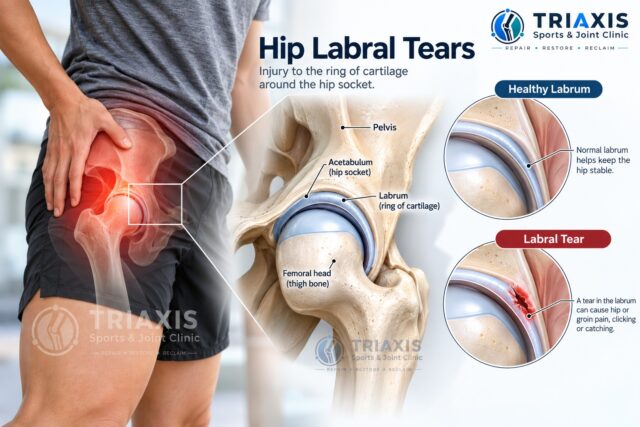
A hip labral tear is an injury to the labrum, the ring of cartilage around the edge of the hip socket. The labrum deepens the socket, helps seal the joint and contributes to its stability. Labral tears may develop after an injury, through repeated loading, or alongside differences in the shape of the hip joint.
The hip is a ball-and-socket joint. The rounded top of the thigh bone, called the femoral head, sits inside a socket in the pelvis called the acetabulum. The labrum runs around the edge of this socket, and most tears occur at the front and top of the rim (the anterosuperior labrum).
A labral injury is one of several possible causes of hip pain. Other causes include muscle and tendon conditions, joint changes, hip impingement and pain referred from the lower back.
Importantly, a labral tear found on a scan does not automatically mean it is responsible for a person’s symptoms. Labral changes are common even in people without hip pain [1], which is why a sports medicine assessment combines the history, examination and imaging rather than relying on a scan report alone.
What does a hip labral tear feel like?
A hip labral tear may cause deep groin or front-of-hip pain, particularly during movements that bend or rotate the hip. Some people notice clicking, catching or stiffness inside the joint. However, symptoms vary, and similar sensations can occur with other hip conditions.
Common symptoms may include:
- Deep groin pain
- Pain at the front of the hip
- Clicking or catching
- Hip stiffness or reduced hip movement
- Pain during twisting or pivoting
- Discomfort during or after sport
- Pain after prolonged sitting
- Difficulty with deep squatting
A 2025 expert consensus of sports medicine physicians and orthopaedic surgeons found that front-of-groin pain, pain with deep hip bending and sharp catching pain during rotation can increase clinical suspicion of a symptomatic labral tear [2].
Where is hip labral tear pain usually felt?
Hip labral tear pain is commonly felt deep in the groin or at the front of the hip. Some people experience discomfort around the side or back of the hip. Pain location alone cannot diagnose a labral tear, because the hip flexor tendons, adductors, gluteal tendons, bursae and lower back can all cause similar symptoms.
Pain may become more noticeable during deep squatting, prolonged sitting, getting out of a low chair or car, twisting, pivoting, lunging, running and changing direction during sport.
If you are unsure where your symptoms may be coming from, the Triaxis guide to Hip Pain: Causes, Symptoms and Treatment Options explains other possible causes of pain around the groin, outer hip, buttock and thigh.
What causes a hip labral tear?
Hip labral tears can develop after a sudden injury or through repeated loading of the hip. They may also occur alongside structural differences such as hip impingement or hip dysplasia, or with gradual changes within the joint over time.
Possible contributing factors include:
- Twisting or pivoting injuries
- Repeated hip rotation and deep hip bending
- Sports-related loading
- Previous hip trauma
- Femoroacetabular impingement
- Hip dysplasia
- Degenerative joint changes
One particularly important relationship is between a labral tear and hip impingement (femoroacetabular impingement or FAI). With hip impingement, the shape of the femoral head, hip socket or both can lead to contact during certain movements, placing additional stress on the labrum and nearby cartilage. However, not everyone with hip impingement develops a symptomatic labral tear, and much of the load on the labrum can be influenced by strength, movement control and training habits, which is exactly what rehabilitation targets.
Which structures make up the hip joint?
The hip is a ball-and-socket joint formed by the pelvis and upper thigh bone. The labrum surrounds the edge of the socket, while cartilage covers the joint surfaces. Muscles, tendons and ligaments around the hip contribute to movement and stability.
Femoral head
The rounded top of the thigh bone that forms the ball of the hip joint.
Acetabulum
The socket within the pelvis.
Labrum
The ring of cartilage around the edge of the acetabulum.
Articular cartilage
Smooth tissue covering the joint surfaces.
Joint capsule and ligaments
Strong bands of tissue that surround the joint and contribute to hip stability.
Muscles and tendons
These include the gluteal muscles, hip flexors (including the iliopsoas) and adductors, which control movement during walking, running and sport.
Nerves
Nerves carry sensation and signals that help control the muscles around the hip and leg.
Because several structures sit close together, pain around the hip or groin does not automatically mean the labrum is injured. Many of these structures can be examined directly with ultrasound during the same consultation.
Who is more likely to develop a hip labral tear?
Hip labral tears can affect active adults and athletes, particularly those involved in repeated hip rotation, pivoting or deep hip bending. Hip shape can also play a role, especially when hip impingement or dysplasia is present.
Factors that may be relevant include previous hip injury, repeated twisting or pivoting, deep hip flexion, high sporting demands, femoroacetabular impingement, hip dysplasia, degenerative changes and repetitive occupational activities. Having one or more of these factors does not mean a person will necessarily develop a symptomatic labral tear.
Which sports are associated with hip labral tears?
Sports involving repeated hip rotation, pivoting, deep bending or rapid changes of direction place greater demands on the hip. However, participating in these sports does not mean a labral tear will develop, and hip pain in an athlete may have many other causes.
Relevant sports and activities include:
- Football and futsal
- Basketball
- Tennis, badminton, pickleball and padel
- Golf
- Running
- Martial arts
- Dance and gymnastics
- Weightlifting

Football, badminton, pickleball and padel involve repeated changes of direction. Golf involves repeated hip rotation. Weightlifting and certain gym exercises involve deeper ranges of hip flexion.
A sports medicine assessment therefore considers what movements the person performs, how training load has changed, and how the hip copes with the specific demands of their sport. This sport-specific view shapes both the rehabilitation plan and the return-to-sport pathway.
How is a hip labral tear diagnosed?
Diagnosis begins with a detailed sports medicine consultation covering the symptoms, their onset and how they affect daily activity or sport. Clinical examination assesses hip movement, strength and the movements that reproduce symptoms. At Triaxis, musculoskeletal ultrasound is usually performed at the same visit, with X-ray or MRI added when they are likely to change management.
Assessment may include:
- Location and pattern of pain
- How and when symptoms started
- Clicking or catching
- Aggravating activities
- Hip range of movement and specific hip tests
- Muscle strength
- Walking and functional movements such as squatting and single-leg control
- Previous hip injuries
- Sporting and occupational demands
- Point-of-care musculoskeletal ultrasound
Symptoms of a labral tear can overlap with hip impingement, tendon conditions and other joint problems, so the assessment also looks carefully for these.
Can ultrasound diagnose a hip labral tear?
Yes, ultrasound has a valuable role. In experienced hands, musculoskeletal ultrasound can assess the front and top of the labrum, where most tears occur, while also checking the joint and surrounding tendons in real time. It is quick, radiation-free and performed in the clinic, and it can guide treatment precisely. MRI or MR arthrogram remains the reference test when a more detailed view of the whole labrum and cartilage is needed.
Musculoskeletal ultrasound uses high-frequency sound waves to produce real-time images of muscles, tendons, ligaments, joints and nerves. Unlike a static scan, it allows the clinician to watch structures move and to correlate findings directly with the patient’s symptoms.
Published studies show that ultrasound can identify anterosuperior labral tears with an overall accuracy of around 84% and a high positive predictive value, and combining several ultrasound signs, such as altered labral echotexture and paralabral cysts, improves its diagnostic performance.
In practice, ultrasound helps in several ways:
| What ultrasound does | Why it matters for a labral tear |
| Visualises the anterosuperior labrum | Assesses the most common site of labral tears |
| Detects joint fluid and paralabral cysts | Supports a joint-related source of pain |
| Examines tendons, bursae and muscles | Identifies or excludes common mimics such as iliopsoas, adductor, rectus femoris or gluteal tendon problems |
| Dynamic, real-time assessment | Allows painful movements, snapping or clicking to be examined as they happen |
| Guides injections precisely | Places diagnostic or therapeutic injections accurately into the joint or target tissue without radiation |
An ultrasound-guided diagnostic injection of local anaesthetic into the hip joint can also help confirm whether pain is arising from inside the joint, an approach recommended by expert consensus when the source of pain is uncertain. The degree of pain relief after an ultrasound-guided hip injection has even been shown to help predict how patients with FAI respond to later treatment.
Do I also need an X-ray or MRI?
Not always. X-rays assess the bones and the shape of the hip, which helps identify impingement, dysplasia or arthritis. MRI or MR arthrogram gives the most detailed view of the labrum and cartilage and is particularly useful when symptoms are not improving or surgical opinion is being considered.
An X-ray may help assess bone structure, joint space, features of hip impingement and hip dysplasia.
MRI provides detailed images of the labrum, cartilage and surrounding soft tissues. In selected cases, an MR arthrogram, which combines MRI with contrast introduced into the joint, may be used for additional detail.
Imaging decisions are tailored to the individual. Many people can begin treatment confidently after clinical assessment and ultrasound, with further imaging added when it is likely to influence management.
Can a hip labral tear improve without surgery?
Yes. Many hip labral tears can be managed successfully without surgery. A pooled analysis of studies in non-arthritic hip-related pain found that more than half of patients reported a satisfactory response to non-surgical treatment, and there is strong evidence supporting hip-specific physiotherapy with activity modification and core strengthening.
The aim of non-surgical care is not to make the tear disappear on a scan. Instead, it aims to calm the irritated joint, reduce the load on the labrum and build a stronger, better-controlled hip that tolerates daily activity and sport.
At Triaxis, non-surgical care combines three elements:
- Accurate diagnosis through sports medicine assessment and ultrasound.
- Structured rehabilitation with physiotherapy and allied health professionals.
- Targeted ultrasound-guided treatments where symptoms are limiting progress.
Current reviews describe a stepwise approach, with non-surgical management first and surgery reserved for appropriately selected patients. Surgical opinion may be considered when symptoms remain significant despite a well-delivered non-surgical programme, or when imaging shows features, such as significant structural deformity or a displaced, unstable tear, that are unlikely to settle without it. Expert consensus also highlights factors such as dysplasia and degenerative changes when weighing up options.
What treatment options are available for a hip labral tear?
Treatment is built around rehabilitation and tailored to the symptoms, hip structure, associated conditions and activity goals. Most plans combine education, activity modification and progressive exercise therapy, supported where appropriate by ultrasound-guided injections such as PRP, HA or prolotherapy.
Education
Understanding the condition helps patients recognise which movements aggravate symptoms and how activities can be adjusted rather than stopped.
Activity modification
This does not mean stopping all exercise. It may involve temporarily adjusting squat depth, running volume, sporting intensity, pivoting, exercise selection and prolonged sitting positions while rehabilitation builds capacity.
Rehabilitation and allied health care
Rehabilitation is the foundation of treatment. Working with physiotherapists, exercise physiologists and strength and conditioning coaches, programmes typically focus on hip and gluteal strength, core and trunk control, single-leg control, movement retraining and a gradual return to running and sport-specific drills. Supervised, hip-specific exercise therapy has the strongest evidence of any non-surgical treatment for hip impingement and associated labral pathology.
Ultrasound-guided injections
For people whose pain is limiting their ability to rehabilitate, an image-guided injection may help settle symptoms and create a window for effective exercise. These treatments are used alongside, not instead of, rehabilitation. Ultrasound guidance ensures accurate placement into the joint or target tissue without radiation exposure [10].
Can PRP, hyaluronic acid or prolotherapy help a hip labral tear?
These injection treatments may help selected patients with persistent symptoms by reducing pain and supporting rehabilitation. They do not reliably “reattach” a torn labrum, and the research is still developing, but they offer useful non-surgical options as part of a combined plan.
| Treatment | What it is | How it may help |
| Platelet-rich plasma (PRP) | A concentrate of platelets prepared from the patient’s own blood, injected into the hip joint under ultrasound guidance | Growth factors and anti-inflammatory proteins may help settle an irritated joint and reduce pain |
| Hyaluronic acid (HA) | A lubricating gel similar to a natural component of joint fluid | May improve joint lubrication and comfort, particularly where early cartilage change is present |
| Prolotherapy | Injection of a dextrose (sugar) solution into the joint, capsule or nearby ligament and tendon attachments | Thought to stimulate a local healing response and reduce pain from the capsule and supporting tissues |
| Diagnostic local anaesthetic, with or without corticosteroid | A precise ultrasound-guided injection into the hip joint | Confirms whether pain is coming from inside the joint and may provide short-term relief |
Studies in hip joint conditions have reported improvements in pain and function following PRP [11], hyaluronic acid [12] [13] and prolotherapy [14], and an ultrasound-guided diagnostic injection helps confirm the source of pain and plan treatment [5]. At Triaxis, these options are discussed as part of shared decision-making, considering the likely source of pain, the stage of rehabilitation, any associated cartilage change and the person’s goals, so that each treatment plan is tailored to the individual.
Treating associated conditions
Hip labral tears frequently coexist with other problems around the hip. Where present, these may be addressed with the wider range of ultrasound-guided treatments available at Triaxis, such as PRP or percutaneous ultrasonic tenotomy (Tenex) for persistent gluteal, hamstring or adductor tendinopathy, shockwave therapy for tendon pain, and nerve hydrodissection where an irritated nerve is contributing to symptoms.
What exercises may help a hip labral tear?
Exercise for a hip labral tear focuses on improving hip and core strength, movement control and tolerance of everyday and sporting activities. There is no single programme that suits everyone, so exercises are individualised and progressed by the rehabilitation team.
Rehabilitation may include:
- Hip and gluteal strengthening
- Core and trunk control
- Balance and single-leg control
- Movement retraining
- Progressive resistance exercise
- Gradual running progression
- Sport-specific exercises
Exercises that repeatedly cause significant symptoms are modified rather than abandoned. For athletes, rehabilitation progresses towards the movements their sport demands, such as running, jumping, pivoting, lunging and changing direction.
What activities can make a hip labral tear feel worse?
Symptoms may become more noticeable during deep hip bending, twisting or rotation. Aggravating activities vary between individuals. The aim is not to avoid movement completely but to identify and temporarily modify the activities that repeatedly reproduce symptoms.
Possible aggravating activities include deep squatting, prolonged or low sitting, getting in and out of a car, pivoting, twisting, lunging, running, kicking and rapid changes of direction. Someone with associated hip impingement may particularly notice symptoms during deep hip flexion and rotational movements.
Activity modification should be individualised rather than based simply on an MRI result.
How long does a hip labral tear take to recover?
There is no single recovery time. Recovery depends on the symptoms, associated hip conditions, physical demands and the treatment approach. Progress is guided by improvements in pain, movement, strength and function rather than a fixed number of weeks.
Many people notice meaningful improvement within the first few months of a well-structured rehabilitation programme, particularly when aggravating loads are adjusted early. Those with longer-standing symptoms or associated hip conditions may need longer, and injection-based treatments may be added if progress stalls.
For athletes, return to sport depends on the ability to run, pivot, squat and change direction comfortably with appropriate strength and control. Return to sport after hip arthroscopy typically takes considerably longer, often around six to eight months [9].
When should I seek medical assessment for hip pain?
Consider a sports medicine assessment if hip or groin pain persists, keeps returning or begins to affect walking, exercise, sport or everyday activities. Early assessment allows rehabilitation to start sooner and gives the best chance of settling symptoms without surgery.
Consider assessment when:
- Groin or hip pain persists
- Pain repeatedly returns during exercise
- Hip movement becomes restricted
- Painful clicking or catching develops
- Symptoms affect walking or sport
- Symptoms follow a sporting injury or fall
More urgent medical attention may be appropriate after significant trauma if there is:
- Severe pain
- Inability to bear weight
- Obvious deformity
- Significant swelling
- New numbness or weakness
- Concern about fracture or dislocation
Not every episode of hip pain represents a labral tear. The broader Triaxis guide to hip pain explains several other conditions that may produce similar symptoms.
How are hip labral tears assessed and treated at Triaxis Sports & Joint Clinic?
At Triaxis Sports & Joint Clinic, care is led by a Consultant in Sports, Exercise & Musculoskeletal Medicine with extensive experience in musculoskeletal ultrasound and ultrasound-guided procedures. Assessment and diagnostic ultrasound usually take place at the same visit, and treatment is planned around rehabilitation, with access to PRP, HA, prolotherapy and other ultrasound-guided treatments where appropriate.
Care may involve:
- Sports and musculoskeletal medicine assessment
- Same-visit diagnostic musculoskeletal ultrasound
- Hip range-of-motion, strength and functional movement testing
- Review of sporting and occupational demands
- Review of existing X-rays or MRI, and further imaging where indicated
- Ultrasound-guided diagnostic injections
- Ultrasound-guided PRP, hyaluronic acid or prolotherapy where clinically appropriate
- Rehabilitation planning with physiotherapy and allied health colleagues
- Return-to-sport planning
- Shared decision-making, with onward surgical opinion where needed
The aim is to identify the true source of symptoms and to provide a clear, non-surgical-first plan that helps people return to the activities and sport they enjoy.
Frequently Asked Questions About Hip Labral Tears
Can a hip labral tear heal on its own?
A labral tear does not need to disappear on imaging for symptoms and function to improve. Many people settle their symptoms with activity modification and structured rehabilitation, sometimes supported by ultrasound-guided injections, without needing surgery. The best approach depends on symptoms, hip structure, associated conditions and activity goals.
Does a hip labral tear always cause pain?
No. Labral tears are common on scans in people with no hip pain at all. Hip and groin symptoms may also come from muscles, tendons, bursae, the joint itself or the lower back, so imaging findings should always be interpreted alongside the symptoms and examination.
Can ultrasound detect a hip labral tear?
Yes, in experienced hands musculoskeletal ultrasound can assess the front and top of the labrum, where most tears occur, and look for related signs such as joint fluid and paralabral cysts. It also checks the surrounding tendons in real time and guides injections accurately. MRI or MR arthrogram may be added when a more detailed view of the whole joint is needed.
Can I exercise with a hip labral tear?
Many people can remain active, with certain movements temporarily modified. Exercise focuses on hip and core strength, movement control and gradually improving tolerance. Rehabilitation is best guided by a physiotherapist or exercise professional working with your sports medicine doctor.
Does a hip labral tear require surgery?
No. Surgery is not automatically required. Many people improve with non-surgical care combining rehabilitation and, where needed, ultrasound-guided injections. Surgical opinion may be considered when symptoms remain significant despite a well-delivered non-surgical programme, or when the hip structure and imaging findings suggest surgery is likely to help.
Can PRP help a hip labral tear?
PRP may help reduce pain and support rehabilitation in selected people with persistent hip joint symptoms. Studies in intra-articular hip conditions show improvements in pain and function for up to a year.
Is hyaluronic acid or prolotherapy an option?
Both may be considered for selected patients. Hyaluronic acid may improve joint comfort, particularly where early cartilage change is present, and prolotherapy may help pain arising from the joint capsule and supporting tissues. Evidence for both is still emerging, and suitability is discussed individually.
How long before I can return to sport?
There is no single timeline. With non-surgical care, return to sport is guided by symptoms, strength and the ability to perform sport-specific movements comfortably. After surgery, rehabilitation and return to sport generally take several months.
About the Author
Dr Dinesh Sirisena is a Consultant in Sports, Exercise & Musculoskeletal Medicine and the author of Ultrasound-Guided Musculoskeletal Procedures in Sports Medicine (Elsevier, 2021). His clinical work focuses on the non-surgical assessment and management of sports injuries and musculoskeletal conditions, including diagnostic musculoskeletal ultrasound and ultrasound-guided treatments.