Disc and Joint Pain: Causes, Symptoms and Treatment Options
Written and medically reviewed by Dr Dinesh Sirisena
Consultant in Sports, Exercise & Musculoskeletal Medicine · Triaxis Sports & Joint Clinic, Farrer Park Hospital, Singapore
In short: Disc and facet joint pain are the two most common structural sources of neck and back pain. Discs act as the spine’s shock absorbers; facet joints guide its movement — and either can become painful through loading, injury or age-related change. Most people improve without surgery, and where pain persists, image-guided facet joint and nerve root injections target the confirmed source precisely.
Key points at a glance
- The intervertebral discs and facet joints are the spine’s most common structural pain sources.
- Disc-related pain is typically central, worse with bending and sitting; facet pain is often one-sided, worse with arching and twisting.
- A disc pressing on a nerve can send pain, tingling or numbness into the arm or leg.
- Disc bulges and facet arthritis are common on scans in pain-free people — imaging must match the examination.
- Most disc and facet pain improves without surgery.
- Image-guided facet joint injections, medial branch blocks and nerve root injections treat the confirmed source precisely.
- Diagnostic blocks can confirm which structure is truly responsible before any bigger decision is made.
What are the discs and facet joints?
Between each pair of vertebrae sits a disc — a shock absorber with a tough outer ring and gel-like centre. Behind them sit the facet joints — small paired joints that guide movement and prevent excessive motion.
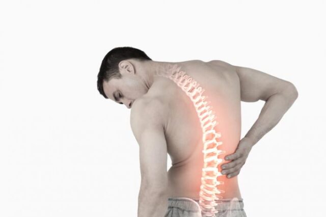
Together they let the spine be both strong and mobile. Each can become painful in its own way: discs through strain of the outer ring or bulging that irritates nearby nerves; facet joints through irritation and arthritis like any other joint in the body.
What does disc pain feel like — and how is facet pain different?
Disc-related pain is typically central or band-like, worse with bending forward, sitting and lifting. Facet joint pain is usually one-sided, worse with arching back, twisting and prolonged standing.
Common patterns include:
- Central low back or neck pain worse with sitting and bending — disc pattern
- One-sided pain worse with arching or twisting — facet pattern
- Sharp catching pain with specific movements
- Pain radiating into the buttock, leg or arm when a nerve root is irritated
- Stiffness after rest that eases with movement — common with facet arthritis
These patterns overlap, and the examination — sometimes supported by a diagnostic block — is what separates them reliably.
What causes disc and facet joint pain?
Loading beyond current capacity, sustained postures and normal age-related change are the usual drivers — a single dramatic injury is the exception, not the rule.
Discs gradually lose water content and height with age, which changes how load transfers to the facet joints — one reason the two problems often coexist. Lifting spikes, prolonged sitting, previous injury and genetics all contribute. Crucially, these changes are frequently painless: they are risk factors, not verdicts.
Who is more likely to develop disc and joint pain?
Adults from their 30s onwards for disc-related pain, and from midlife onwards for facet joint pain — with manual workers, desk workers and rotation-sport athletes most represented.
Genetics play a larger role in disc degeneration than most people expect, which is another reason self-blame about posture is usually misplaced.
How is disc and facet joint pain diagnosed?
Diagnosis combines the pain pattern, movement testing and neurological examination — and, where a precise answer matters, a diagnostic image-guided block that temporarily numbs the suspected structure.
The diagnostic block is an underused tool: if numbing a facet joint or nerve root abolishes the pain, the source is confirmed with a confidence no scan can provide. This protects patients from treatments — including surgery — aimed at the wrong structure.
Do I need an MRI?
Only if it will change management: persistent nerve symptoms, red flags, or planning a targeted procedure. Most disc and facet pain is diagnosed and managed clinically.
Disc bulges appear on MRI in a large proportion of pain-free adults, and facet arthritis is near-universal with age. A scan finding only matters when it matches the clinical picture — which is why the examination leads and the scan follows.
How is disc and facet joint pain treated?
Active rehabilitation is the foundation, and most people improve with it. Image-guided procedures precisely target the confirmed source where pain persists.
A complete treatment programme may include:
- Education and graded activity — movement is treatment, and fear of movement worsens outcomes
- A progressive strengthening programme matched to your pain pattern (flexion- or extension-biased)
- Image-guided facet joint injections or medial branch blocks for confirmed facet pain
- Image-guided nerve root (transforaminal/epidural) injections where a disc is irritating a nerve
- Diagnostic blocks to confirm the pain source before any larger decision
- Prolotherapy or PRP for selected cases as part of a complete treatment programme
Every injection is performed with continuous image guidance so the medication reaches exactly the joint or nerve responsible — and always alongside rehabilitation, which is what delivers the durable result.
What exercises help?
Progressive strengthening of the trunk, hips and back — matched to your movement preference — plus regular walking. Consistency matters more than any single exercise.
Disc-pattern pain often prefers extension-based early work; facet-pattern pain often prefers flexion-biased work. Getting this match right early accelerates recovery — another dividend of an accurate assessment.
How long does it take to improve?
Most episodes improve substantially over two to six weeks; nerve root pain from a disc typically settles over six to twelve weeks as the disc bulge naturally regresses.
That last point surprises many patients: the majority of disc herniations shrink on their own over months — the body resorbs them. This is a large part of why surgery is rarely needed.
Do I need surgery for a disc problem?
Rarely. Surgery is indicated for progressive weakness, cauda equina syndrome, or severe nerve pain that has failed comprehensive non-surgical care including image-guided injections.
For back pain alone — without those features — the evidence does not favour surgery over structured non-surgical care. If disc surgery has been recommended without a trial of image-guided treatment, a second opinion is a reasonable step; many patients on surgical waiting lists improve before their date arrives.
When should I seek medical assessment?
Seek assessment if pain persists beyond a few weeks or radiates into a limb — and urgently for saddle numbness, bladder or bowel changes, or progressive leg weakness.
Those urgent features are rare but time-critical, and they are the reason radiating pain should be assessed rather than watched indefinitely.
Assessment at Triaxis Sports & Joint Clinic
Triaxis Sports & Joint Clinic at Farrer Park Hospital provides assessment for spinal pain, disc-related symptoms and facet joint conditions.
Assessment includes a full musculoskeletal and neurological examination, honest interpretation of any imaging, and open discussion of the full range of options — rehabilitation programmes, diagnostic blocks, and image-guided facet joint and nerve root injections — so you understand every option before deciding on a plan.
Frequently asked questions
Can a disc bulge heal on its own?
Yes — most disc herniations regress naturally over months as the body resorbs the bulged material, and symptoms typically improve ahead of the scan. This is a central reason surgery is rarely needed.
How do I know if my pain is from the disc or the facet joint?
The pattern gives strong clues — disc pain is typically central and worse with bending and sitting; facet pain one-sided and worse with arching and twisting. Where certainty matters, an image-guided diagnostic block confirms the source definitively.
What is a medial branch block?
A precise, image-guided injection that numbs the small nerves supplying a facet joint. It serves as both a diagnostic test — confirming the joint as the pain source — and a treatment that settles pain so rehabilitation can progress.
Is sitting bad for my discs?
Sitting loads the discs more than standing, but the real problem is unbroken hours of it. Regular position changes and a strengthening programme let most people sit comfortably through a working day.
Will my facet arthritis keep getting worse?
Facet changes on scans progress with age, but pain does not have to follow — strength, mobility and load management control symptoms for most people, with image-guided injections in reserve for flare-ups.
Disc or joint pain in your neck or back that isn’t settling?
Most disc and facet joint pain improves without surgery. Dr Sirisena offers thorough assessment, rehabilitation planning, diagnostic blocks and precise image-guided facet and nerve root injections — as a first port of call or a second opinion.
Triaxis Sports & Joint Clinic, Farrer Park Hospital · WhatsApp +65 8889 3594 · hello@triaxisclinic.com · Repair · Restore · Reclaim
About the author
Written and medically reviewed by Dr Dinesh Sirisena, Consultant in Sports, Exercise & Musculoskeletal Medicine, and author of Ultrasound-Guided Musculoskeletal Procedures in Sports Medicine (Elsevier, 2021). Dr Sirisena focuses on the non-surgical and image-guided treatment of musculoskeletal and sports injuries.