
Sports Ultrasound Imaging: Uses, Benefits and What to Expect
Written and medically reviewed by
Dr Dinesh Sirisena
Consultant in Sports, Exercise & Musculoskeletal Medicine
Triaxis Sports & Joint Clinic
Sports ultrasound imaging is a form of musculoskeletal ultrasound used to assess selected muscles, tendons, ligaments, joints, nerves and other soft tissues, particularly in the context of sports and exercise injuries. It uses sound waves rather than ionising radiation and can provide real-time images during movement. However, it is not needed for every injury, and MRI or X-ray may sometimes be more appropriate.
What is sports ultrasound imaging?
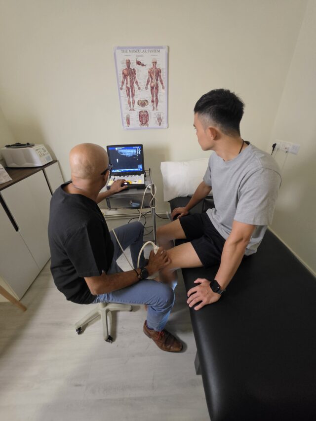
Sports ultrasound imaging is musculoskeletal ultrasound used in the assessment of sports-related and exercise-related conditions. It can produce real-time images of selected muscles, tendons, ligaments, joints, nerves and soft tissues, helping clinicians investigate certain injuries and understand how some structures behave during movement.
A small handheld device called a transducer is placed on the skin with ultrasound gel. High-frequency sound waves travel into the body and return to the probe. A computer then converts these signals into images.
Because the images appear immediately, clinicians can sometimes examine selected structures while the patient moves.
This is one reason ultrasound can be useful in Sports Medicine, where symptoms may appear only during a particular movement.
It can therefore help answer a specific question and aid your clinical assessment.
Is sports ultrasound the same as musculoskeletal ultrasound?
Yes, the terms overlap considerably. Musculoskeletal ultrasound describes ultrasound imaging of muscles, tendons, ligaments, joints, nerves and related soft tissues. “Sports ultrasound” usually refers to the same imaging technique when it is used within Sports Medicine or for exercise-related injuries and movement problems.
The main difference is the clinical setting rather than the technology.
For example, sports ultrasound may be used when assessing:
- A runner with Achilles tendon pain
- A tennis player with elbow symptoms
- A swimmer with shoulder pain
- A footballer with a muscle injury
- A badminton player with an ankle injury
The usefulness of ultrasound still depends on the structure involved and the suspected diagnosis.
What can sports ultrasound imaging detect?
Sports ultrasound can help assess selected tendon injuries, muscle tears, ligament problems, joint fluid, soft tissue lumps and certain nerve conditions. However, while it cannot evaluate every structure equally well, such as deeper tissues or areas hidden by bone, it may be useful in guiding treatments for these areas.
Common examples include:
- Rotator cuff tendon problems
- Biceps tendon conditions
- Tennis elbow
- Golfer’s elbow
- Achilles tendinopathy
- Patellar tendon problems
- Muscle strains and tears
- Selected ligament injuries
- Ganglion cysts
- Selected peripheral nerve problems
- Fluid within certain joints or bursae
Tendon tears, muscle abnormalities, ligament sprains, joint or bursal fluid and nerve entrapment are also among other conditions that can be assessed using musculoskeletal ultrasound.
Which body areas can sports ultrasound examine?
Sports ultrasound can assess many superficial structures around the shoulder, elbow, wrist, hand, hip region, knee, ankle and foot. It is particularly useful when the structure of interest lies relatively close to the skin. However, while it cannot evaluate every structure equally well, such as deeper tissues or areas hidden by bone, it may be useful in guiding treatments for these areas.
Common examples include:
- Shoulder: rotator cuff tendons, biceps tendon and selected surrounding structures.
- Elbow: common extensor and flexor tendons, selected ligaments and nerves.
- Wrist and hand: tendons, superficial nerves and soft-tissue lumps.
- Hip region: selected tendons and superficial soft tissues.
- Knee: quadriceps tendon, patellar tendon, selected ligaments and superficial structures.
- Ankle and foot: Achilles tendon, peroneal tendons, other ankle tendons, plantar fascia and selected ligaments.
The scan should be chosen based on the suspected condition, not simply the body part that hurts.
Can sports ultrasound show muscles, tendons, ligaments and nerves?
Sports ultrasound can produce images of selected muscles, tendons, ligaments, peripheral nerves and joints. It can also show some of these structures while they move, which may provide useful information when symptoms occur during a specific sporting movement or joint position.
This may help assess:
- Tendon movement
- Muscle contraction
- Tendon subluxation
- Selected nerve movement
- Ligament behaviour
- Joint-related movement
The choice of investigation will be discussed with you by your clinician and in experienced hands, ultrasound can be an excellent first line investigation in the clinic setting.
Why is dynamic ultrasound useful in sports injuries?
Dynamic ultrasound allows a clinician to assess certain musculoskeletal structures while the patient moves. This can be useful when a tendon, joint or nerve behaves differently during motion, or when symptoms occur only in a specific position that may not be reproduced on a static scan.
For example, a patient may be asked to:
- Rotate the shoulder
- Flex or extend the wrist
- Move the ankle
- Contract a muscle
- Perform a movement linked to their symptoms
Ultrasound can show soft-tissue movement in real time, including tendons and joints.
Dynamic assessment is an additional feature of ultrasound, and sometimes this makes it a preferable investigation over a static MRI.
Which sports are commonly associated with injuries assessed by ultrasound?
Sports ultrasound may be considered for injuries arising in running, football, tennis, badminton, golf, swimming, cricket, rugby, cycling, weightlifting and other activities. The type of injury depends on the sport, training demands, movement pattern and body region involved rather than on the sport alone.
Examples include:
- Running: Achilles tendon, calf muscle and selected ankle injuries
- Tennis: elbow, shoulder and wrist tendon problems
- Badminton: ankle, knee, shoulder and elbow injuries
- Golf: elbow, wrist and shoulder conditions
- Swimming: shoulder tendon problems
- Football and rugby: muscle and selected ligament injuries
- Weightlifting: shoulder, elbow and tendon-related problems
- Cricket: shoulder, elbow and muscle injuries
Different sports may encounter different injuries and it is important that a thorough clinical assessment is undertaken as well as relevant investigations.
Is sports ultrasound better than MRI?
Ultrasound can assess selected superficial soft tissues in real time and during movement, while MRI can show deeper tissues, bone marrow, cartilage and internal joint structures that ultrasound may not visualise well. The most appropriate test depends on the clinical question. However, ultrasound enables more dynamic assessments and rapid comparison with the other side.
Ultrasound may be useful when assessing:
- Superficial tendons
- Selected ligament injuries
- Muscle injuries
- Peripheral nerves
- Dynamic movement
MRI may be preferable for:
- Deep joint structures
- Bone marrow
- Cartilage
- Internal joint injuries
- Structures hidden behind bone
While ultrasound cannot evaluate every structure equally well, such as deeper tissues or areas hidden by bone, it may be useful in guiding treatments for these areas.
What is the difference between sports ultrasound, MRI and X-ray?
Sports ultrasound, MRI and X-ray provide different types of information. X-rays mainly assess bones and certain joint changes. MRI provides detailed views of deeper soft tissues and internal joint structures. Sports ultrasound provides real-time imaging of selected superficial muscles, tendons, ligaments and nerves without ionising radiation.
| Imaging method | Common role |
| X-ray | Bones, fractures, alignment and some joint changes |
| MRI | Deeper soft tissues, cartilage, bone marrow and internal joint structures |
| Sports ultrasound | Selected muscles, tendons, ligaments, nerves and superficial joints |
| Dynamic ultrasound | Observing selected structures during movement |
With ultrasound available in the clinic setting, it enables rapid assessment of injuries in real time and with dynamic capabilities.
Do all sports injuries need ultrasound imaging?
All sports injuries should initially be evaluated with a thorough medical history and physical examination before using imaging. Sports ultrasound may be considered when it is likely to clarify the suspected injury, assess a particular structure or influence management. Ultrasound can help determine what may be contributing to your underling pain.
A clinician may first examine:
- How the injury occurred
- The location of symptoms
- Swelling or bruising
- Strength
- Joint movement
- Stability
- Functional movements
- Training history
If these findings suggest a condition that ultrasound can assess well, imaging may then be considered.
Can sports ultrasound help diagnose tendon injuries?
Sports ultrasound can be useful for assessing selected tendon conditions, including tendinopathy and some partial or complete tendon tears. It can also show tendon movement dynamically. Together with your clinical and physical assessment, ultrasound can help shed further light on the condition.
Common tendons assessed with ultrasound may include:
- Rotator cuff tendons
- Biceps tendon
- Common elbow tendons
- Patellar tendon
- Achilles tendon
- Peroneal tendons
- Selected wrist and hand tendons
Rotator cuff and Achilles tendon tears or tendinitis are among the most common musculoskeletal conditions assessed with sports ultrasound.
Can sports ultrasound help assess ligament injuries?
Ultrasound can assess selected superficial ligaments, particularly around areas such as the ankle, elbow and wrist. Dynamic examination may also help in some cases. However, while deeper ligaments or complex joint injuries may require MRI or other imaging, ultrasound may be useful in guiding treatments for these areas.
For example, ultrasound may have a role in selected ankle ligament injuries.
Current ACR guidance lists ultrasound as potentially appropriate in some cases of suspected chronic ankle instability or ligament injury after radiographs, while MRI may also be appropriate.
The choice should therefore depend on the injury pattern rather than a general preference for one test.
Can sports ultrasound detect muscle tears?
Ultrasound can help assess selected muscle injuries, including some strains, tears, fluid collections and other soft-tissue changes. It may also allow the muscle to be viewed while contracting. While, MRI may sometimes be considered when the injury is deep or difficult to characterise adequately, ultrasound may be useful in guiding treatments for these areas.
Muscle injuries are common in sports involving:
- Sprinting
- Kicking
- Jumping
- Sudden acceleration
- Rapid changes of direction
Examples include football, rugby, athletics and court sports.
Imaging should be considered together with the history and physical examination and can help guide further management.
Can sports ultrasound guide injections or procedures?
Ultrasound can be used to visualise selected anatomical structures during certain musculoskeletal procedures. This can help the clinician identify the intended target and surrounding tissues in real time.
Depending on the individual’s condition and clinical assessment, certain procedures may be considered as part of a broader management plan.
Possible uses of ultrasound guidance may include selected:
- Joint procedures
- Tendon-related procedures
- Bursa procedures
- Other soft-tissue interventions
Procedures should be undertaken after a thorough clinical and physical assessment. Sometimes they can also have a diagnostic role where symptoms or the diagnosis is unclear.
It will usually be combined with rehabilitation, exercise and load management.
How does sports ultrasound fit into rehabilitation planning?
Sports ultrasound may provide additional information about a selected structure, but rehabilitation should not be based on the scan alone. Symptoms, strength, mobility, movement control, sporting demands and clinical findings all contribute to exercise planning and decisions about returning to activity.
A rehabilitation programme may include:
- Education
- Activity modification
- Progressive strengthening
- Mobility exercises
- Load management
- Balance and movement training
- Sport-specific rehabilitation
- Gradual return to training
Two patients with similar ultrasound findings may require different rehabilitation plans because their symptoms, goals and physical demands differ.
What happens during a sports ultrasound examination?
During a sports ultrasound examination, gel is placed on the skin and an ultrasound probe is moved over the area being assessed. The clinician may view the structure from different angles and may ask the patient to move, contract a muscle or reproduce a particular motion during the scan.
Most musculoskeletal ultrasound examinations require little or no preparation.
During the scan, the clinician may:
- Identify the relevant anatomy
- Examine the structure from several angles
- Compare different positions
- Observe movement
- Assess selected blood-flow patterns using Doppler where appropriate
- Relate findings to the physical examination
Standard diagnostic ultrasound does not use X-ray radiation.
When should I seek assessment for a sports injury?
Consider medical assessment when a sports injury causes persistent pain, significant swelling, weakness, instability, loss of function or repeated difficulty returning to activity. Assessment is also appropriate when the diagnosis is unclear or symptoms do not improve as expected. The clinician can then decide whether ultrasound or another investigation is useful.
More urgent assessment may be appropriate when there is:
- Significant trauma
- Obvious deformity
- Inability to bear weight or use the limb
- Sudden major weakness
- Severe swelling
- New numbness
- Fever or feeling significantly unwell
- A hot and markedly swollen joint
Imaging should be undertaken to support your clinical and physical assessment.
How is sports ultrasound imaging used at Triaxis Sports & Joint Clinic?
At Triaxis Sports & Joint Clinic, sports ultrasound may be considered as part of the clinical assessment of selected sports and musculoskeletal conditions. Its use depends on the patient’s symptoms, examination findings and the anatomical structure involved. Where appropriate, ultrasound findings can support patient education, management decisions and rehabilitation planning.
Assessment may include:
- Sports and injury history
- Musculoskeletal examination
- Movement and functional assessment
- Sports ultrasound where appropriate
- Discussion of imaging findings
- Patient education
- Rehabilitation planning
- Shared decision-making
Where an X-ray, MRI or another investigation is more suitable, that can be considered according to the clinical findings.
This factual approach is also more consistent with Singapore healthcare advertising requirements, which emphasise accurate claims and avoidance of exaggerated statements that could encourage unnecessary treatment.
Frequently Asked Questions About Sports Ultrasound Imaging
Is sports ultrasound safe?
Diagnostic sports ultrasound does not use ionising radiation. Instead, it uses high-frequency sound waves to create images of musculoskeletal structures. Standard diagnostic ultrasound has no known harmful effects when used appropriately. As with other investigations, it should still be performed when there is a clinical reason rather than routinely for every sports injury.
Can sports ultrasound detect a tendon tear?
Ultrasound can identify selected tendon abnormalities, including some partial or complete tears. Its usefulness depends on the tendon involved, the depth of the structure and the quality of the clinical question. Some injuries may require MRI for additional information, particularly when deeper tissues or internal joint structures need assessment.
Can ultrasound diagnose an ankle sprain?
Ultrasound can assess selected ankle ligaments and tendons, but it is not necessary for every ankle sprain. The clinical history and physical examination are usually the starting point. Depending on the suspected injury, radiographs, ultrasound or MRI may be considered. For selected chronic ligament or tendon problems, ultrasound may be appropriate.
Can sports ultrasound show inflammation?
Ultrasound can show selected changes in tendons, joints, bursae and other soft tissues, while Doppler ultrasound may provide information about blood flow in some situations. The scan needs to be interpreted alongside the patient’s symptoms and physical examination.
Can sports ultrasound assess nerve problems?
Musculoskeletal ultrasound can visualise selected peripheral nerves and may help assess certain nerve entrapments or structural changes. Symptoms such as numbness, tingling or weakness can have several causes, so a neurological examination and sometimes additional tests may be required.
Is sports ultrasound painful?
The scan itself is usually non-invasive. Some tenderness may occur when the probe presses over an already painful or injured area. If ultrasound is being used to guide a procedure, that is different from a diagnostic scan. Patients should therefore understand whether the appointment is for imaging, a procedure or both.
Will I need an MRI after sports ultrasound?
Ultrasound may answer some clinical questions adequately, while MRI may be more suitable for others. Further imaging may be considered when deeper tissues, cartilage, bone marrow or internal joint structures need assessment, or when symptoms remain unexplained. The decision should depend on the clinical findings rather than follow automatically after ultrasound.
Can sports ultrasound be performed during a Sports Medicine consultation?
Where clinically appropriate and available, sports ultrasound may form part of a Sports Medicine or Musculoskeletal Medicine assessment. It can provide real-time information about selected soft tissues and may complement the examination. Ultrasound aims to complement your medical history, physical examination and broader assessment of movements and sporting demands.
About the Author, Dr Dinesh Sirisena is a Consultant in Sports, Exercise & Musculoskeletal Medicine. His clinical work includes assessment and management of sports injuries and musculoskeletal conditions involving muscles, tendons, ligaments, joints and related structures.
Medical Disclaimer
This page provides general educational information and does not replace an individual medical consultation. Sports and musculoskeletal injuries can have different causes, and not every condition requires ultrasound or other imaging. Appropriate investigations and management depend on the individual’s symptoms, medical history and clinical assessment.